. 2021 Apr 23;10(5):985.
doi: 10.3390/cells10050985.

Anti-Tumor Effects of MAPK-Dependent Tumor-Selective Oncolytic Vaccinia Virus Armed with CD/UPRT against Pancreatic Ductal Adenocarcinoma in Mice

Affiliations

Anti-Tumor Effects of MAPK-Dependent Tumor-Selective Oncolytic Vaccinia Virus Armed with CD/UPRT against Pancreatic Ductal Adenocarcinoma in Mice

Hajime Kurosaki et al. Cells. .

Abstract

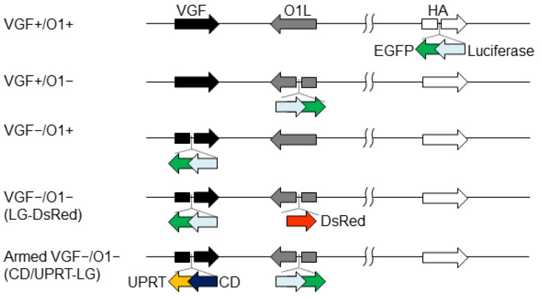
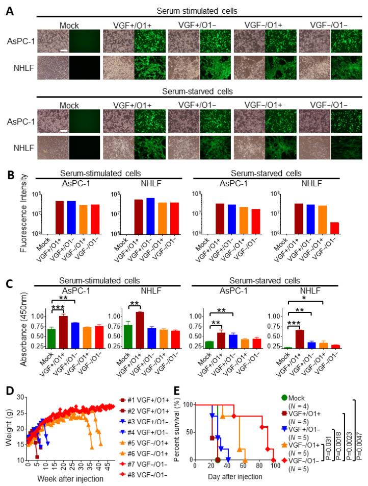
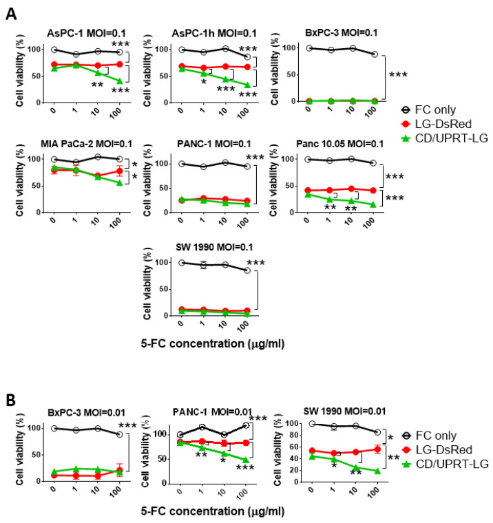
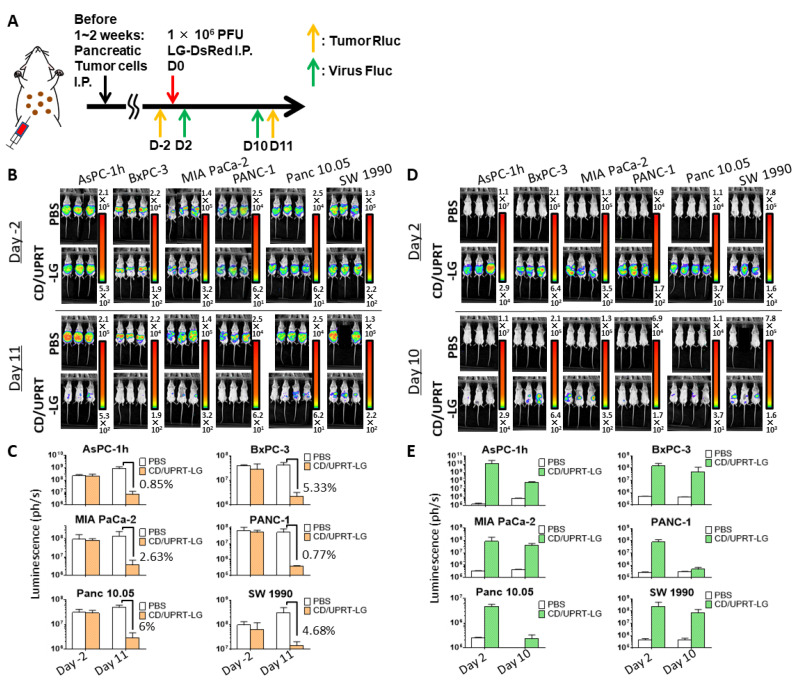
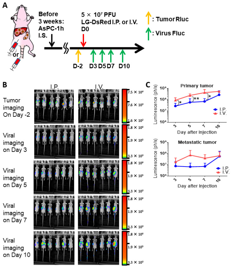
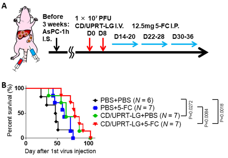
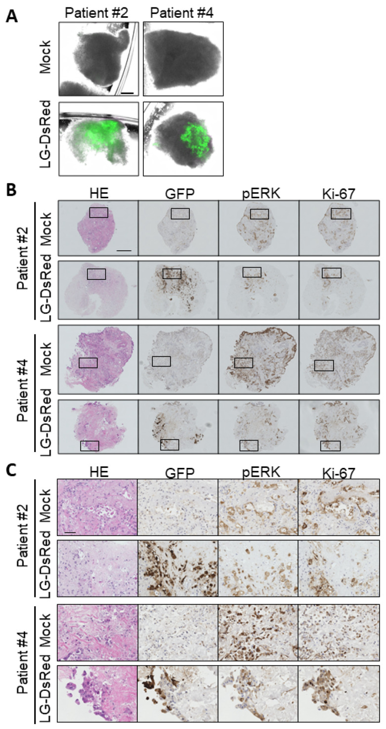
Engineered vaccinia virus serves as an oncolytic virus for cancer virotherapy. We evaluated the oncolytic characteristics of VGF- and O1-deleted recombinant mitogen-activated protein kinase (MAPK)-dependent vaccinia virus (MDRVV). We found that compared with viruses with the deletion of either gene alone, MDRVV is more attenuated in normal cells and can replicate in cancer cells that exhibit constitutive ERK1/2 activation in the MAPK pathway. We armed MDRVV with a bifunctional fusion gene encoding cytosine deaminase and uracil phosphoribosyltransferase (CD/UPRT), which converts 5-fluorocytosine (5-FC) into chemotherapeutic agents, and evaluated its oncolytic activity alone or in combination with 5-FC in human pancreatic cancer cell lines, tumor mouse models of peritoneal dissemination and liver metastasis, and ex vivo-infected live pancreatic cancer patient-derived tissues. CD/UPRT-armed MDRVV alone could efficiently eliminate pancreatic cancers, and its antitumor effects were partially enhanced in combination with 5-FC in vitro and in vivo. Moreover, the replication of MDRVV was detected in tumor cells of patient-derived, surgically resected tissues, which showed enlarged nuclei and high expression of pERK1/2 and Ki-67, and not in stromal cells. Our findings suggest that systemic injections of CD/UPRT-armed MDRVV alone or in combination with 5-FC are promising therapeutic strategies for pancreatic ductal adenocarcinoma.

Keywords: CD/UPRT; MAPK pathway; antitumor effects; oncolytic vaccinia virus; pancreatic cancer; prodrug 5-FC.


Conflict of interest statement

The authors declare no conflict of interest.

Figures

Figure 1
Figure 1
Recombinant vaccinia viruses genetically engineered by the deletion of viral genes and expression of transgenes. Vaccinia growth factor (VGF)- and/or O1-deleted viruses were generated through the insertion of a gene cassette expressing luciferase-fused EGFP (LG) and/or DsRed into the VGF and O1 gene loci. VGF- and O1-deleted armed virus was developed via the insertion of a gene cassette expressing cytosine deaminase-fused uracil phosphoribosyl transferase (CD/UPRT) and LG into the VGF and O1 gene loci, respectively. VGF- and O1-intact control viruses were constructed through the insertion of a gene cassette expressing LG into the HA gene loci.
Figure 2
Figure 2
Vaccinia growth factor (VGF)- and/or O1-deleted mitogen-activated protein kinase-dependent recombinant vaccinia viruses (MDRVVs) are highly attenuated in normal cells but retain their therapeutic replication. The human pancreatic cancer cell line (AsPC-1) or normal human lung fibroblast (NHLF) cells were infected with each recombinant virus at a multiplicity of infection (MOI) of 1 under serum stimulation or starvation conditions, and were cultured for 30 h. (A) Bright-field (left) and EGFP (right) representative images of AsPC-1 and NHLF cells are shown. Scale bar, 500 μm. (B) The fluorescence intensity in EGFP images of (A) was measured using Hybrid Cell Count (n = 1). (C) The levels of phosphorylated ERK1/2 (pERK1/2) were evaluated. Data are presented as the mean ± SD (n = 3). * p < 0.05, ** p < 0.01, *** p < 0.001 (one-way ANOVA and the Bonferroni test). (D) SCID mice were injected intraperitoneally with 1 × 10⁶ plaque-forming units (PFUs) of each recombinant virus, and changes in body weight were observed for 48 weeks (n = 2). (E) BxPC-3 cells stably expressing Renilla luciferase were injected intraperitoneally into SCID mice on day 0. On day 8, the mice received a single intraperitoneal injection of each recombinant virus (1 × 10⁶ PFU). Survival curves of mice were generated by Kaplan–Meier analysis (n = 4–5). p values were derived by log-rank test.
Figure 3
Figure 3
Cell viability tests show cytotoxicity with mitogen-activated protein kinase-dependent recombinant vaccinia virus (MDRVV) alone or in combination with armed MDRVV and 5- fluorocytosine (5-FC) in human pancreatic ductal adenocarcinoma cell lines. (A) Pancreatic cancer cell lines were infected with each recombinant virus at a multiplicity of infection (MOI) of 0.1, and 48 h later. Cells were treated with 0, 1, 10, or 100 μg/mL of 5-FC. Cell viabilities were determined 120 h after virus infection. (B) BxPC-3, PANC-1, and SW 1990 cells were infected with each recombinant virus at an MOI of 0.01 and combined with 5-FC as described in (A). Data are presented as the mean ± SD (n = 3). * p < 0.05, ** p < 0.01, *** p < 0.001 (two-tailed unpaired t-test).
Figure 4
Figure 4
Mitogen-activated protein kinase-dependent recombinant vaccinia virus (MDRVV) (CD/UPRT-LG) efficiently eliminates peritoneally disseminated tumors and its replication decreases following tumor elimination in mouse tumors derived from human pancreatic ductal adenocarcinoma cell lines. (A) Schematic representation of the study design and timeline of tumor growth and viral distribution. Severe combined immunodeficiency mice with peritoneally disseminated six pancreatic cancer cell lines expressing Renilla luciferase (Rluc) were intraperitoneally injected with CD/UPRT-LG (1 × 10⁶ PFU). (B) Tumor Rluc luminescence was detected on day 2 before and on day 11 after virus inoculation. One mouse bearing peritoneally disseminated SW 1990 xenografts died due to tumor burden 9 days after PBS treatment. (C) Quantification of tumor Rluc luminescence was determined from (B). (D) Viral firefly luciferase (Fluc) luminescence was determined on days 2 and 10 after virus inoculation. One mouse bearing peritoneally disseminated SW 1990 xenografts died due to tumor burden 9 days after PBS treatment. (E) Quantification of viral Fluc luminescence was determined from (D). The data are presented as the mean + SD (n = 2–4).
Figure 5
Figure 5
Comparison of intraperitoneal (I.P.) or intravenous (I.V.) injections of mitogen-activated protein kinase-dependent recombinant vaccinia virus (MDRVV) (LG-DsRed) in a mouse model harboring liver metastasis caused by human pancreatic ductal adenocarcinoma cells. (A) Schematic representation of the study design and timeline of tumor growth and viral distribution. (B) AsPC-1 CD44v9-high cells expressing Renilla Luciferase (Rluc; AsPC-1h) were injected intrasplenically into athymic nude mice and liver metastasis was detected via tumor Rluc luminescence on day 2 before viral treatment. Mice were injected intraperitoneally or intravenously with 5 × 10⁷ PFU of MDRVV (LG-DsRed). Viral replication was detected on days 3, 5, 7, and 10 via firefly luciferase (Fluc) luminescence. (C) Quantification of viral Fluc luminescence in primary splenic and metastatic liver sites was determined from (B). The data are presented as the mean ± SD (n = 6). * p < 0.05, ** p < 0.01 (two-tailed unpaired t-test).
Figure 6
Figure 6
Combination treatment with mitogen-activated protein kinase-dependent recombinant vaccinia virus (MDRVV) (CD/UPRT-LG) and 5-fluorocytosine (5-FC) increases the therapeutic effect in a mouse model harboring liver metastasis caused by human pancreatic ductal adenocarcinoma cells. (A) Schematic representation of the study design and timeline of tumor growth and viral distribution. (B) The nude mice with liver metastasis were intravenously injected with 1 × 10⁷ PFU of MDRVV (CD/UPRT-LG) on days 0 and 8. Next, 12.5 mg 5-FC was injected intraperitoneally into the mice every day from days 14 to 36, except on days 21 and 29. Survival curves of mice were generated by Kaplan–Meier analysis (n = 6–7) and p values were determined using the log-rank test.
Figure 7
Figure 7
Mitogen-activated protein kinase-dependent recombinant vaccinia virus (MDRVV) achieves tumor-selective replication in tissue cultures from pancreatic ductal adenocarcinoma (PDAC) patients. (A) Minced pancreatic tissue samples derived from human pancreatic cancer patients were infected with 1 × 10⁶ plaque-forming units (PFU) of LG-DsRed. Viral GFP was photographed at 120 h after infection (scale bar: 300 μm). (B) Immunohistochemical staining of human tissue samples in (A). Tissues were stained with hematoxylin and eosin, an anti-GFP antibody, an anti-phosphorylated p44/42 MAPK protein (Erk1/2) antibody, or anti-Ki-67 (scale bar: 300 μm). (C) Extended images of those marked with squares in (B) (scale bar: 50 μm).

Similar articles

Cited by

References

    1. Kim M. Replicating Poxviruses for Human Cancer Therapy. J. Microbiol. 2015;53:209–218. doi: 10.1007/s12275-015-5041-4. - DOI - PubMed
    1. Guo Z.S., Lu B., Guo Z., Giehl E., Feist M., Dai E., Liu W., Storkus W.J., He Y., Liu Z., et al. Vaccinia Virus-mediated Cancer Immunotherapy: Cancer Vaccines and Oncolytics. J. Immunother. Cancer. 2019;7:6. doi: 10.1186/s40425-018-0495-7. - DOI - PMC - PubMed
    1. Kim J.H., Oh J.Y., Park B.H., Lee D.E., Kim J.S., Park H.E., Roh M.S., Je J.E., Yoon J.H., Thorne S.H., et al. Systemic Armed Oncolytic and Immunologic Therapy for Cancer With JX-594, a Targeted Poxvirus Expressing GM-CSF. Mol. Ther. 2006;14:361–370. doi: 10.1016/j.ymthe.2006.05.008. - DOI - PubMed
    1. McCart J.A., Ward J.M., Lee J., Hu Y., Alexander H.R., Libutti S.K., Moss B., Bartlett D.L. Systemic Cancer Therapy with a Tumor-Selective Vaccinia Virus Mutant Lacking Thymidine Kinase and Vaccinia Growth Factor Genes. Cancer Res. 2001;61:8751–8757. - PubMed
    1. Twardzik D.R., Brown J.P., Ranchalis J.E., Todaro G.J., Moss B. Vaccinia Virus-infected Cells Release a Novel Polypeptide Functionally Related to Transforming and Epidermal Growth Factors. Proc. Natl. Acad. Sci. USA. 1985;82:5300–5304. doi: 10.1073/pnas.82.16.5300. - DOI - PMC - PubMed

Publication types

MeSH terms