

Radiomics and Magnetic Resonance Imaging of Rectal Cancer: From Engineering to Clinical Practice
- PMID: 33922483
- PMCID: PMC8146913
- DOI: 10.3390/diagnostics11050756
Radiomics and Magnetic Resonance Imaging of Rectal Cancer: From Engineering to Clinical Practice
Abstract
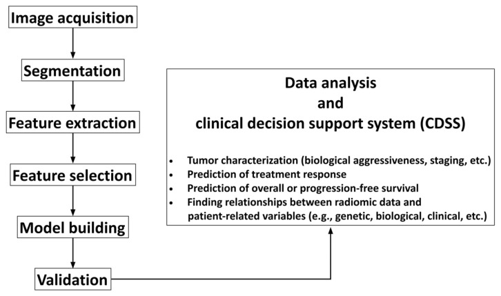
While cross-sectional imaging has seen continuous progress and plays an undiscussed pivotal role in the diagnostic management and treatment planning of patients with rectal cancer, a largely unmet need remains for improved staging accuracy, assessment of treatment response and prediction of individual patient outcome. Moreover, the increasing availability of target therapies has called for developing reliable diagnostic tools for identifying potential responders and optimizing overall treatment strategy on a personalized basis. Radiomics has emerged as a promising, still fully evolving research topic, which could harness the power of modern computer technology to generate quantitative information from imaging datasets based on advanced data-driven biomathematical models, potentially providing an added value to conventional imaging for improved patient management. The present study aimed to illustrate the contribution that current radiomics methods applied to magnetic resonance imaging can offer to managing patients with rectal cancer.
Keywords: deep learning; magnetic resonance imaging; neoadjuvant chemoradiation therapy; personalized medicine; radiomics; rectal cancer; surgery.
Conflict of interest statement
The authors declare no conflict of interest.
Figures


References
-
- DʼSouza N., de Neree Tot Babberich M.P.M., d’Hoore A., Tiret E., Xynos E., Beets-Tan R.G.H., Nagtegaal I.D., Blomqvist L., Holm T., Glimelius B., et al. Definition of the rectum: An international, expert-based Delphi consensus. Ann. Surg. 2019;270:955–959. doi: 10.1097/SLA.0000000000003251. - DOI - PubMed
-
- Beets-Tan R.G.H., Lambregts D.M.J., Maas M., Bipat S., Barbaro B., Curvo-Semedo L., Fenlon H.M., Gollub M.J., Gourtsoyianni S., Halligan S., et al. Magnetic resonance imaging for clinical management of rectal cancer: Updated recommendations from the 2016 European Society of Gastrointestinal and Abdominal Radiology (ESGAR) consensus meeting. Eur. Radiol. 2018;28:1465–1475. doi: 10.1007/s00330-017-5026-2. - DOI - PMC - PubMed
-
- Gollub M.J., Arya S., Beets-Tan R.G., de Prisco G., Gonen M., Jhaveri K., Kassam Z., Kaur H., Kim D., Knezevic A., et al. Use of magnetic resonance imaging in rectal cancer patients: Society of Abdominal Radiology (SAR) rectal cancer disease-focused panel (DFP) recommendations 2017. Abdom. Radiol. 2018;43:2893–2902. doi: 10.1007/s00261-018-1642-9. - DOI - PubMed
