Accuracy and Reproducibility of Facial Measurements of Digital Photographs and Wrapped Cone Beam Computed Tomography (CBCT) Photographs
- PMID: 33922543
- PMCID: PMC8147051
- DOI: 10.3390/diagnostics11050757
Accuracy and Reproducibility of Facial Measurements of Digital Photographs and Wrapped Cone Beam Computed Tomography (CBCT) Photographs
Abstract
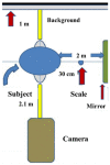
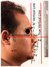
The study sought to assess whether the soft tissue facial profile measurements of direct Cone Beam Computed Tomography (CBCT) and wrapped CBCT images of non-standardized facial photographs are accurate compared to the standardized digital photographs. In this cross-sectional study, 60 patients with an age range of 18-30 years, who were indicated for CBCT, were enrolled. Two facial photographs were taken per patient: standardized and random (non-standardized). The non-standardized ones were wrapped with the CBCT images. The most used soft tissue facial profile landmarks/parameters (linear and angular) were measured on direct soft tissue three-dimensional (3D) images and on the photographs wrapped over the 3D-CBCT images, and then compared to the standardized photographs. The reliability analysis was performed using concordance correlation coefficients (CCC) and depicted graphically using Bland-Altman plots. Most of the linear and angular measurements showed high reliability (0.91 to 0.998). Nevertheless, four soft tissue measurements were unreliable; namely, posterior gonial angle (0.085 and 0.11 for wrapped and direct CBCT soft tissue, respectively), mandibular plane angle (0.006 and 0.0016 for wrapped and direct CBCT soft tissue, respectively), posterior facial height (0.63 and 0.62 for wrapped and direct CBCT soft tissue, respectively) and total soft tissue facial convexity (0.52 for both wrapped and direct CBCT soft tissue, respectively). The soft tissue facial profile measurements from either the direct 3D-CBCT images or the wrapped CBCT images of non-standardized frontal photographs were accurate, and can be used to analyze most of the soft tissue facial profile measurements.
Keywords: cone beam computed tomography; facial photographs; standardized photograph; wrapped photographs.
Conflict of interest statement
The authors declare no conflict of interest.
Figures


References
-
- Zyman P., Etienne J.-M. Recording and communicating shade with digital photography: Concepts and considerations. Pract. Proced. Aesthetic Dent. 2002;14:49–51. - PubMed
LinkOut - more resources
Full Text Sources

