. 2021 Apr 23;11(5):1219.
doi: 10.3390/ani11051219.

A Prototype Skin Substitute, Made of Recycled Marine Collagen, Improves the Skin Regeneration of Sheep

Affiliations

A Prototype Skin Substitute, Made of Recycled Marine Collagen, Improves the Skin Regeneration of Sheep

Luca Melotti et al. Animals (Basel). .

Abstract

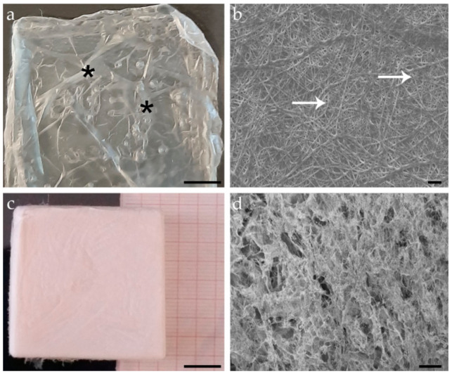
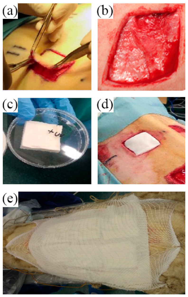
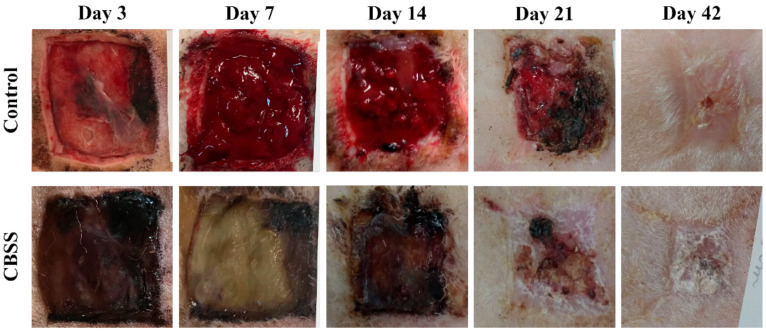
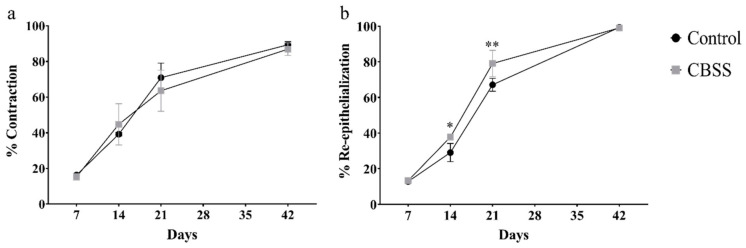
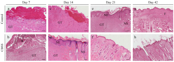
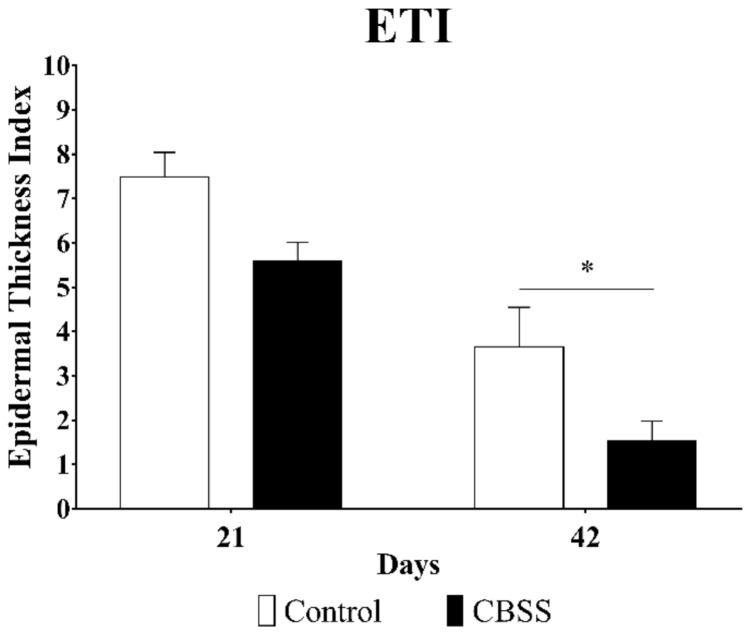
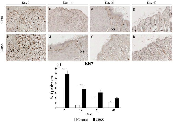
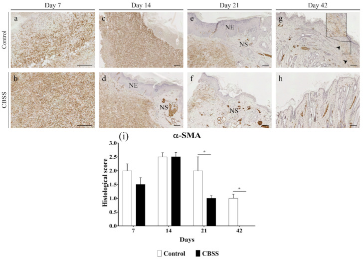
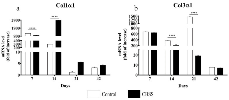
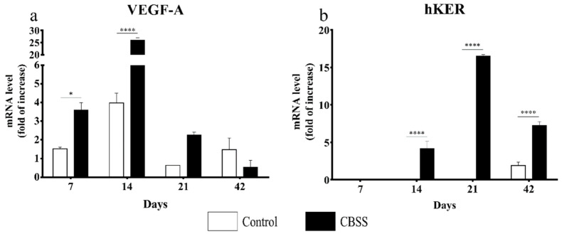
Skin wound healing is a complex and dynamic process that aims to restore lesioned tissues. Collagen-based skin substitutes are a promising treatment to promote wound healing by mimicking the native skin structure. Recently, collagen from marine organisms has gained interest as a source for producing biomaterials for skin regenerative strategies. This preliminary study aimed to describe the application of a collagen-based skin-like scaffold (CBSS), manufactured with collagen extracted from sea urchin food waste, to treat experimental skin wounds in a large animal. The wound-healing process was assessed over different time points by the means of clinical, histopathological, and molecular analysis. The CBSS treatment improved wound re-epithelialization along with cell proliferation, gene expression of growth factors (VEGF-A), and development of skin adnexa throughout the healing process. Furthermore, it regulated the gene expression of collagen type I and III, thus enhancing the maturation of the granulation tissue into a mature dermis without any signs of scarring as observed in untreated wounds. The observed results (reduced inflammation, better re-epithelialization, proper development of mature dermis and skin adnexa) suggest that sea urchin-derived CBSS is a promising biomaterial for skin wound healing in a "blue biotechnologies" perspective for animals of Veterinary interest.

Keywords: 3D scaffolds; biomaterials; circular economy; innovative therapies; marine collagen; regenerative medicine; sea urchin; skin substitute; tissue engineering; wound healing.


Conflict of interest statement

The authors declare no conflict of interest.

Figures

Figure 1
Figure 1
Examples of 2D membranes and 3D scaffolds of sea urchin-derived collagen. (a) Top view of a 2D membrane (light microscopy). Asterisks mark macroscopic folds of the thin 2D membrane. (b) Micrograph of a 2D membrane where the random distribution of the single collagen fibrils (arrows) in the two-dimensional network is visible (scanning electron microscopy). (c) Top view of a 3D scaffold (light microscopy). (d) Micrograph of a 3D scaffold where the porous microstructure of the biomaterial is detectable (scanning electron microscopy). Scalebar: (a) = 500 µm; (b) = 2 µm; (c) = 1 cm; (d) = 200 µm.
Figure 2
Figure 2
Representative images of the surgery procedure and biomaterial implantation. (a) Skin full-thickness removal by using surgical scissors for detaching the dermis from the subcutis; (b) macroscopic appearance of the wound after surgery; (c) 3D scaffold after UV sterilization and before implantation; (d) 3D scaffold implantation in the wound, the biomaterial was directly placed onto the wound bed; (e) representative figure of the bandages applied to the back of each sheep after the surgery.
Figure 3
Figure 3
Representative images of the skin ulcers during wound healing.
Figure 4
Figure 4
(a) Percentage of contraction. (b) Percentage of re-epithelialization. Data are shown as mean ± SEM. * p < 0.05; ** p < 0.01.
Figure 5
Figure 5
Histopathological microphotographs of skin biopsies at different time points after wounding: control and CBSS-treated wounds comparison. (a,b) Skin wounds at 7 days, in the CBSS-treated wounds (b) is possible to appreciate the presence of the 3D scaffold; (c,d) wounds at 14 days, treated wounds started to show a neoepidermis (NE, characterized by an hyperplastic appearance) and skin adnexa; (e,f) wounds at 21 days after wounding; (g,h) wounds at 42 days. GT = granulation tissue; NE = neoepidermis; NS = neoskin; F = fibrosis; asterisk = 3D sponge-like scaffold. Scalebar = 200 μm.
Figure 6
Figure 6
Epidermal thickness index (ETI) at 21 and 42 days respect to unwounded skin. Data are shown as mean ± SEM. * p < 0.05.
Figure 7
Figure 7
Immunohistochemistry microphotographs for Ki67 immunolabeling. Wounds are showed at (a,b) 7 days, (c,d) 14 days, (e,f) 21 days, and (g,h) 42 days. (i) Quantitative analysis of the percentage of positive area of each sample at 7, 14, 21, and 42 days. Arrowhead = active proliferating keratinocytes in the epidermal basal layer; NE = neoepidermis; NS = neoskin. Scalebar = 200 μm. (i) Data are expressed as mean ± SEM. Statistical differences were measured between the two experimental groups at the same time-point. **** p < 0.0001.
Figure 8
Figure 8
Immunohistochemistry microphotographs for α-SMA immunostaining. Wounds are showed at (a,b) 7 days, (c,d) 14 days, (e,f) 21 days, and (g,h) 42 days. (i) Semi-quantitative analysis based on the score for presence and orientation of myofibroblasts in wounds at 7, 14, 21, and 42 days. Arrowhead = myofibroblasts in the mature dermis; NE = neoepidermis; NS = neoskin. Scalebar = 200 μm; inset = higher magnification of the dermal fibrosis. (i) Data are expressed as mean ± SEM. Statistical differences were measured between the two experimental groups at the same time point * p < 0.05.
Figure 9
Figure 9
Gene expression analysis for collagen genes involved in skin wound healing. (a) Relative expression of the collagen type I (Col1α1) gene and (b) collagen type III (Col3α1) gene at 7, 14, 21, and 42 days after wounding in control and CBSS-treated wounds. Data are shown as mean ± SEM. Unwounded skin was used as the calibrator sample. Statistical differences were measured between the two experimental groups at the same time point. **** p < 0.0001.
Figure 10
Figure 10
Gene expression analysis for VEGF and hKER genes (a) Relative expression of the vascular endothelial growth factor A (VEGF-A) gene and (b) hair-Keratin (hKER) gene at 7, 14, 21, and 42 days after wounding in control and CBSS-treated wounds. Data are shown as mean ± SEM. Unwounded skin was used as the calibrator sample. Statistical differences were measured between the two experimental groups at the same time point. * p < 0.05; **** p < 0.0001.

References

    1. Theoret C.L. The pathophysiology of wound repair. Vet. Clin. N. Am. Equine Pract. 2005;21:1–13. doi: 10.1016/j.cveq.2004.11.001. - DOI - PubMed
    1. Frykfors von Hekkel A.K., Pegram C., Halfacree Z.J. Thoracic dog bite wounds in dogs: A retrospective study of 123 cases (2003–2016) Vet. Surg. 2020;49:694–703. doi: 10.1111/vsu.13402. - DOI - PubMed
    1. Rothe K., Tsokos M., Handrick W. Animal and Human Bite Wounds. Dtsch. Arztebl. Int. 2015;112:433–443. doi: 10.3238/arztebl.2015.0433. - DOI - PMC - PubMed
    1. Cerqueira M.T., Pirraco R.P., Marques A.P. Stem Cells in Skin Wound Healing: Are We There Yet? Adv. Wound Care. 2016;5:164–175. doi: 10.1089/wound.2014.0607. - DOI - PMC - PubMed
    1. Sparks H.D., Sigaeva T., Tarraf S., Mandla S., Pope H., Hee O., Di Martino E.S., Biernaskie J., Radisic M., Scott W.M. Biomechanics of Wound Healing in an Equine Limb Model: Effect of Location and Treatment with a Peptide-Modified Collagen-Chitosan Hydrogel. ACS Biomater. Sci. Eng. 2021;7:265–278. doi: 10.1021/acsbiomaterials.0c01431. - DOI - PubMed