. 2021 Apr 23;13(5):1423.
doi: 10.3390/nu13051423.

Anti-Inflammatory and Anti-Oxidative Synergistic Effect of Vitamin D and Nutritional Complex on Retinal Pigment Epithelial and Endothelial Cell Lines against Age-Related Macular Degeneration

Affiliations

Anti-Inflammatory and Anti-Oxidative Synergistic Effect of Vitamin D and Nutritional Complex on Retinal Pigment Epithelial and Endothelial Cell Lines against Age-Related Macular Degeneration

Maria Hernandez et al. Nutrients. .

Abstract

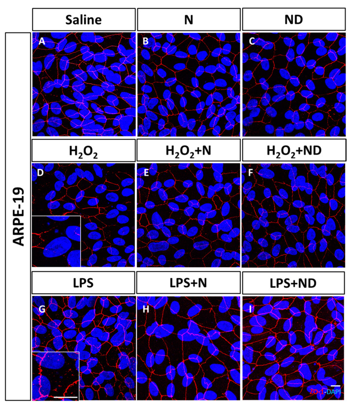
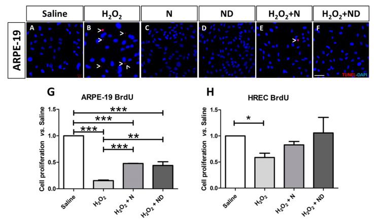
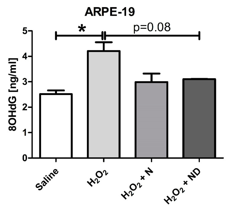
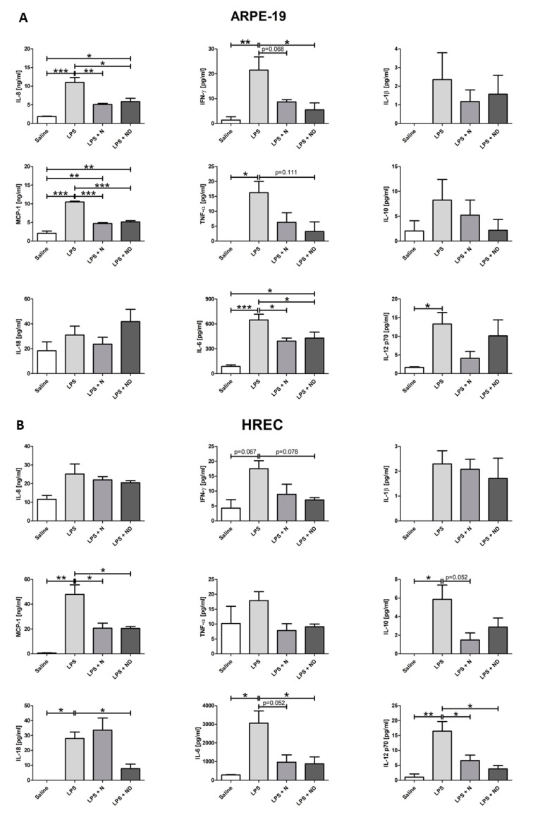
Age-related macular degeneration (AMD) is a multifactorial disease of the retina featured by dysfunction of retinal pigmented epithelial (RPE) and loss of photoreceptor cells under oxidative stress and inflammatory conditions. Vitamin D and antioxidants have beneficial effects against retinal degenerative diseases, such as AMD. We investigated the impact of associating vitamin D (ND) with a nutritional antioxidant complex (Nutrof Total®; N) on oxidative stress and inflammation-like induced conditions by H2O2 and LPS, respectively, in human retinal epithelial (ARPE-19) and human retinal endothelial (HREC) cells. Application of either N or ND treatments to H2O2-induced media in ARPE-19 cells counteracted late apoptosis, attenuated oxidative DNA damage, and increased cell proliferation. Significant reduction in the expression levels of MCP1, IL-8, and IL6 cytokines was observed following application of either N or ND treatments under LPS-induced conditions in ARPE-19 cells and in MCP-1 and IL12p70 cytokine levels in HREC cells. ND and not N revealed significant downregulation of IFNγ in ARPE-19 cells, and of IL-6 and IL-18 in HREC cells. In conclusion, adding vitamin D to Nutrof Total® protects in a synergistic way against oxidative and inflammatory stress-induced conditions in retinal epithelial and endothelial cells.

Keywords: AMD; inflammation; nutritional complex; oxidative stress; retina; vitamin D.


Conflict of interest statement

A.G.-L. is consultant for Bayer, Novartis, Allergan, Thea, and Roche. The rest of the authors declare no conflict of interest. The funders had no role in the design of the study; in the collection, analyses, or interpretation of data; in the writing of the manuscript; or in the decision to publish the results.

Figures

Figure 1
Figure 1
Junctional integrity of ARPE-19 cells evaluated by ZO-1 (red) fluorescence under a confocal imaging system. Application of N and ND treatments (62.34 μg/mL each) did not affect tight junctions, cell integrity, and structure (B,C) compared to saline (A). Addition of H2O2 (2h; 1600 μM) and LPS (24 h; 20 μg/mL) to induce oxidative stress and inflammatory-like conditions, respectively, damaged tight junctions (D,G), while incubation with N and ND treatments during the last hour of the induction recovered the altered structure (E,F,H,I). Nuclei were labelled with DAPI (blue). Scale bar: 20 µm.
Figure 2
Figure 2
Late apoptosis assessed in ARPE-19 cells by TUNEL labelling and imaged under a confocal microscope. Application of N and ND treatments (62.34 μg/mL) to the media (C,D) showed similar results to saline (A). Addition of H2O2 (2 h; 600 μM) to induce oxidative stress increased TUNEL-positive-stained ARPE-19 cells (B; red), while TUNEL labelling was absent by application of N and ND treatments (62.34 μg/mL each) during the last hour of the induction (E,F). Nuclei were labelled with DAPI (blue). Scale bar: 50 µm. Cell proliferation assay was performed in both ARPE-19 and HREC cells (G,H), n = 3. BrdU expression levels were significantly reduced in both cell lines following oxidative stress-induced conditions by 1000 μM H2O2 for 2h. This effect was significantly recovered in ARPE-19 cells by application of N (p < 0.001) and ND (p < 0.01) treatments (62.34 μg/mL each) during the last hour of the induction, while a strong tendency towards significance was observed in HREC cells (* p < 0.05, ** p < 0.001, *** p < 0.001).
Figure 3
Figure 3
Oxidative damage to DNA was analyzed by 8OHdG expression levels for ARPE-19 cells measured by ELISA (n = 3). Addition of H2O2 (2 h; 1000 μM) to induce oxidative stress conditions significantly (p < 0.05) increased 8OHdG levels. Application of N and ND treatments (62.34 μg/mL each) during the last hour of the induction time were able to reduce 8OHdG levels in the media, but not to significant levels (p = 0.08). (* p < 0.05).
Figure 4
Figure 4
Multiple cytokine expression assays in ARPE-19 and HREC cells (n = 3). (A) Expression levels of IL-8, MCP-1, TNF-α, IFNγ, IL-6, and IL-12p70 were significantly increased (p < 0.05) following addition of LPS (24 h; 20 μg/mL) to induce inflammatory-like conditions in ARPE cells. IL-8, MCP-1, and IL-6 expression levels were significantly reduced (p < 0.05) following application of N and ND (62.34 μg/mL each) during the last hour of the induction time. Addition of ND in LPS-induced media was able to significantly decrease (p < 0.05) the expression levels of IFNγ and attenuate the expression of TNFα (p = 0.111), while N showed a strong tendency to reduce IFN-γ (p = 0.068). (B) Expression levels of MCP-1, IL-10, IL-18, IL-6, and IL-12p70 were significantly increased (p < 0.05) after addition of LPS to induce inflammatory-like conditions (24h; 50 μg/mL) in HREC cells. Expression levels of MCP-1 and IL-12p70 were significantly reduced (p < 0.05) following application of N and ND (62.34 μg/mL each) during the last hour of the induction time. Addition of ND, and not N, in LPS-induced media significantly decreased IL-6 and IL-18 expression levels (p < 0.05), while there was a tendency to downregulate IFNγ (p = 0.078). Addition of N attenuated expression of IL-6 and IL-10 (p = 0.052). All cytokines are expressed in pg/mL except for MCP-1 and IL-8 that were expressed as ng/mL. (* p < 0.05, ** p < 0.001, *** p < 0.001).

Similar articles

Cited by

References

    1. Lim L.S., Mitchell P., Seddon J.M., Holz F.G., Wong T.Y. Age-related macular degeneration. Lancet. 2012;379:1728–1738. doi: 10.1016/S0140-6736(12)60282-7. - DOI - PubMed
    1. Gheorghe A., Mahdi L., Musat O. Age-related macular degeneration. Rom. J. Ophthalmol. 2015;59:74–77. - PMC - PubMed
    1. Mitchell P., Liew G., Gopinath B., Wong T.Y. Age-related macular degeneration. Lancet. 2018;392:1147–1159. doi: 10.1016/S0140-6736(18)31550-2. - DOI - PubMed
    1. Klein R., Cruickshanks K.J., Nash S.D., Krantz E.M., Nieto F.J., Huang G.H., Pankow J.S., Klein B.E. The prevalence of age-related macular degeneration and associated risk factors. Arch. Ophthalmol. 2010;128:750–758. doi: 10.1001/archophthalmol.2010.92. - DOI - PMC - PubMed
    1. Ferris F.L., 3rd, Wilkinson C.P., Bird A., Chakravarthy U., Chew E., Csaky K., Sadda S.R. Clinical classification of age-related macular degeneration. Ophthalmology. 2013;120:844–851. doi: 10.1016/j.ophtha.2012.10.036. - DOI - PMC - PubMed

MeSH terms

LinkOut - more resources