

The Relationship between Pulmonary Damage and Peripheral Vascular Manifestations in Systemic Sclerosis Patients
- PMID: 33922710
- PMCID: PMC8145021
- DOI: 10.3390/ph14050403
The Relationship between Pulmonary Damage and Peripheral Vascular Manifestations in Systemic Sclerosis Patients
Abstract
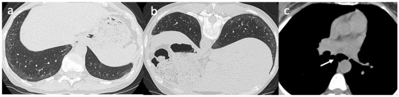
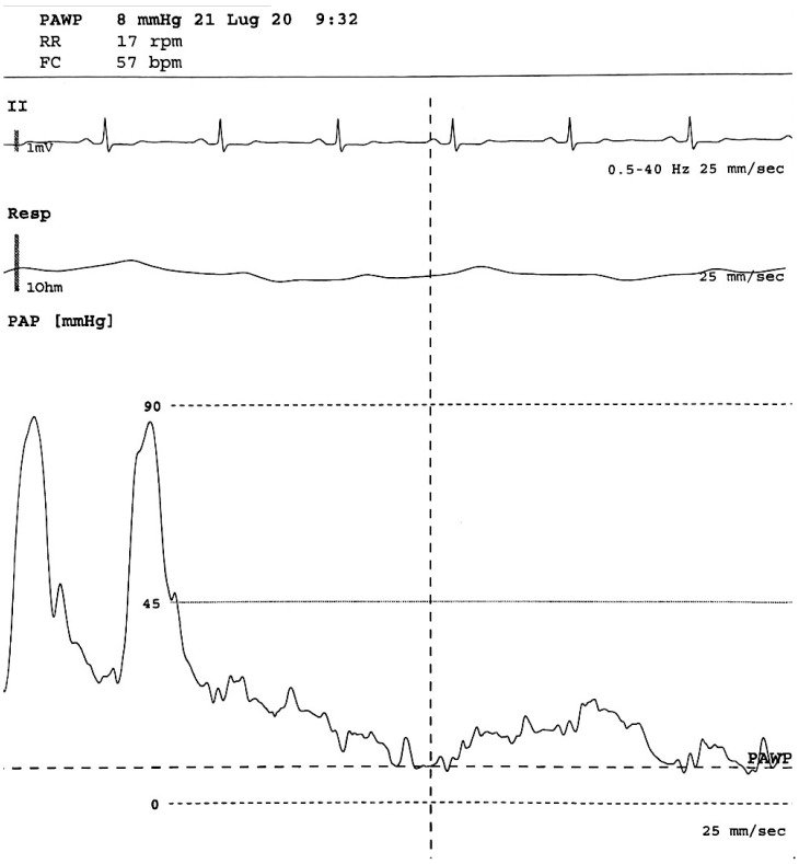
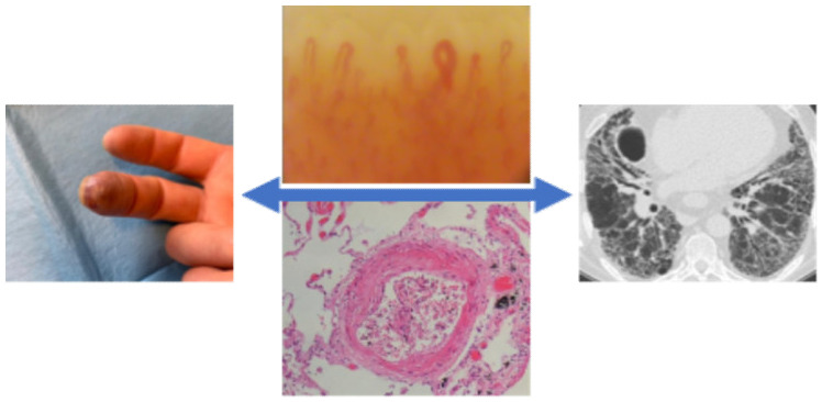
Systemic sclerosis (SSc) is an autoimmune disease, characterized by the presence of generalized vasculopathy and tissue fibrosis. Collagen vascular disorder in SSc is due to fibroblast and endothelial cell dysfunctions. This leads to collagen overproduction, vascular impairment and immune system abnormalities and, in the last stage, multi-organ damage. Thus, to avoid organ damage, which has a poor prognosis, all patients should be carefully evaluated and followed. This is particularly important in the initial disease phase, so as to facilitate early identification of any organ involvement and to allow for appropriate therapy. Pulmonary disease in SSc mainly involves interstitial lung disease (ILD) and pulmonary arterial hypertension (PAH). High-resolution computed tomography (HRCT) and pulmonary function tests (PFT) have been proposed to monitor parenchymal damage. Although transthoracic echocardiography is the most commonly used screening tool for PAH in SSc patients, definitive diagnosis necessitates confirmation by right heart catheterization (RHC). Moreover, some studies have demonstrated that nailfold videocapillaroscopy (NVC) provides an accurate evaluation of the microvascular damage in SSc and is able to predict internal organ involvement, such as lung impairment. This review provides an overview of the correlation between lung damage and microvascular involvement in SSc patients.
Keywords: interstitial lung disease; microvascular involvement; nailfold capillaroscopy; pulmonary arterial hypertension; pulmonary involvement; systemic sclerosis.
Conflict of interest statement
The authors declare that there are no conflict of interest.
Figures
References
-
- Varga J., Trojanowska M., Kuwana M. Pathogenesis of systemic sclerosis: Recent insights of molecular and cellular mechanisms and therapeutic opportunities. J. Scleroderma Relat. Disord. 2017;2:137–152. doi: 10.5301/jsrd.5000249. - DOI
-
- Ruaro B., Soldano S., Smith V., Paolino S., Contini P., Montagna P., Pizzorni C., Casabella A., Tardito S., Sulli A., et al. Correlation between circulating fibrocytes and dermal thickness in limited cutaneous systemic sclerosis patients: A pilot study. Rheumatol. Int. 2019;39:1369–1376. doi: 10.1007/s00296-019-04315-7. - DOI - PubMed
-
- Bruni C., Frech T., Manetti M., Rossi F.W., Furst D.E., De Paulis A., Rivellese F., Guiducci S., Matucci-Cerinic M., Bellando-Randone S. Vascular leaking, a pivotal and early pathogenetic event in systemic sclerosis: Should the door be closed? Front. Immunol. 2018;9:2045. doi: 10.3389/fimmu.2018.02045. - DOI - PMC - PubMed
Publication types
LinkOut - more resources
Full Text Sources
