Review
. 2021 Apr 23;22(9):4434.
doi: 10.3390/ijms22094434.

Fabry Disease and the Heart: A Comprehensive Review

Affiliations
Review

Fabry Disease and the Heart: A Comprehensive Review

Olga Azevedo et al. Int J Mol Sci. .

Abstract

Fabry disease (FD) is an X-linked lysosomal storage disorder caused by mutations of the GLA gene that result in a deficiency of the enzymatic activity of α-galactosidase A and consequent accumulation of glycosphingolipids in body fluids and lysosomes of the cells throughout the body. GB3 accumulation occurs in virtually all cardiac cells (cardiomyocytes, conduction system cells, fibroblasts, and endothelial and smooth muscle vascular cells), ultimately leading to ventricular hypertrophy and fibrosis, heart failure, valve disease, angina, dysrhythmias, cardiac conduction abnormalities, and sudden death. Despite available therapies and supportive treatment, cardiac involvement carries a major prognostic impact, representing the main cause of death in FD. In the last years, knowledge has substantially evolved on the pathophysiological mechanisms leading to cardiac damage, the natural history of cardiac manifestations, the late-onset phenotypes with predominant cardiac involvement, the early markers of cardiac damage, the role of multimodality cardiac imaging on the diagnosis, management and follow-up of Fabry patients, and the cardiac efficacy of available therapies. Herein, we provide a comprehensive and integrated review on the cardiac involvement of FD, at the pathophysiological, anatomopathological, laboratory, imaging, and clinical levels, as well as on the diagnosis and management of cardiac manifestations, their supportive treatment, and the cardiac efficacy of specific therapies, such as enzyme replacement therapy and migalastat.

Keywords: Fabry disease; cardiomyopathy; enzyme replacement therapy; heart; migalastat.


Conflict of interest statement

O.A. and G.M.-M. have received educational/research grants from Shire Human Genetic Therapies/Takeda and travel/accommodation support for conferences from Shire Human Genetic Therapies/Takeda, Amicus and Sanofi Genzyme. M.F.G. has received travel/accommodation support for conferences from Shire Human Genetic Therapies, Amicus, and Sanofi Genzyme. C.F. has received travel/accommodation support for conferences from Shire Human Genetic Therapies/Takeda, Amicus, and Sanofi Genzyme. The remaining authors declare no conflicts of interest.

Figures

Figure 1
Figure 1
Left ventricular hypertrophy (LVH) secondary to Fabry disease (FD). Transthoracic echocardiogram of a 70-year-old male Fabry patient showing a severe symmetrical LVH with prominent papillary muscles in parasternal long-axis (A), four-chambers (B) and short-axis (C) views.
Figure 2
Figure 2
Cardiac magnetic resonance imaging (MRI) (3.0 Tesla) in four Fabry patients, illustrating different stages of myocardial involvement by the disease. (A) Cine (balanced steady-state free precession sequence) image at the basal left ventricular (LV) short-axis slice; (B) Late gadolinium enhancement (LGE) at the basal LV short-axis slice; (C) Native T1 mapping (precontrast) performed using a modified Look-Locker inversion (MOLLI) recovery sequence at the basal LV short-axis slice (the resulting pixel-by-pixel T1 color maps were displayed using a customized lookup table, in which normal myocardium was green, increasing T1 was yellow and red, and decreasing T1 was blue); and (D) T2 mapping (fast low angle shot (FLASH)) at basal LV short-axis slice (the resulting pixel-by-pixel T2 color maps were displayed using a customized lookup table, in which normal myocardium was purple and increasing T2 was red and yellow). Patient 1: A 26-year-old female without LVH or LGE, presenting normal values of T1 (1265 ± 68 ms at the basal septum) and T2 (41.76 ± 5.40 ms at the basal septum); Patient 2: A 41-year-old male without LVH or LGE, presenting low T1 (1118 ± 40 ms) and normal T2 (40.93 ± 5.80 ms) values at the basal septum; Patient 3: A 76-year-old female with LVH (LV mass 83 g/m2, maximum wall thickness 19 mm at the basal septum) and diffuse LGE in the basal segment of the inferolateral wall, who presents low T1 (1093 ± 36 ms) at the basal septum and T1 pseudonormalization particularly at the inferolateral wall (1276 ± 59 ms), where a mild increase in T2 values (49.10 ± 2.30 ms) was also observed; Patient 4: A 69-year-old male patient with LVH (LV mass 123 g/m2, maximum wall thickness 18 mm at the septum) and diffuse and extensive LGE in the inferolateral wall, who presents areas of T1 pseudonormalization but also areas of T1 increase, such as in the inferolateral wall (1425 ± 144 ms), where T2 values (64.44 ± 8.56 ms) are also increased.
Figure 3
Figure 3
Strain echocardiography for the detection of LGE. (A) Transthoracic echocardiogram with 2D-strain analysis by speckle tracking showing reduction of longitudinal strain, particularly in the inferolateral, inferior and inferoseptal walls and apex, in a 73-year-old male Fabry patient; (BE) Cardiac MRI (3.0 Tesla) with LGE in short-axis slices, from the LV base to the apex, showing fibrosis in the same regions where the reduction in longitudinal strain was more pronounced.
Figure 4
Figure 4
Cardiac MRI (3.0 Tesla) in a Fabry patient with advanced cardiomyopathy. Cine (balanced steady-state free precession sequence) images at the basal LV short-axis slice (A) and four-chamber view (C) showing massive and asymmetrical LVH (maximal thickness 30. mm at the septum) with thinning of the posterior wall (2 mm). LGE at the basal LV short-axis slice (B) and four-chamber view (D) showing fibrosis of the inferior and inferolateral walls and apex and focal fibrosis in the septum, where the LVH is more pronounced.
Figure 5
Figure 5
Apical sparing pattern of global longitudinal strain (GLS) in a Fabry patient. Transthoracic echocardiogram of a 48-year-old male Fabry patient showing symmetrical LVH in a four-chamber view (A) and 2 D-strain analysis by speckle tracking revealing an apical sparing pattern of GLS (B).
Figure 6
Figure 6
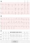
Electrocardiographic findings in FD. (A) Short PR interval in a 34-year-old male, (B) bifascicular and first-degree AV blocks and electrocardiographic criteria of LVH in a 72-year-old male, and (C) non-sustained VT in 24 h-Holter monitoring of a 76-year-old male.

References

    1. Desnick R.J., Ioannou Y.A., Eng C.M. Alpha-galactosidase A deficiency: Fabry disease. In: Scriver C.R., Beaudet A.L., Sly W.S., Valle D., Childs B., Kinzler K.W., Vogelstein B., editors. The Metabolic and Molecular Bases of Inherited Disease. McGraw Hill; New York, NY, USA: 2001. pp. 3733–3774.
    1. Germain D.P. Fabry disease. Orphanet J. Rare Dis. 2010;5:30. doi: 10.1186/1750-1172-5-30. - DOI - PMC - PubMed
    1. Arends M., Wanner C., Hughes D., Mehta A., Oder D., Watkinson O.T., Elliott P.M., Linthorst G.E., Wijburg F.A., Biegstraaten M., et al. Characterization of Classical and Nonclassical Fabry Disease: A Multicenter Study. J. Am. Soc. Nephrol. 2017;28:1631–1641. doi: 10.1681/ASN.2016090964. - DOI - PMC - PubMed
    1. Azevedo O., Gal A., Faria R., Gaspar P., Miltenberger-Miltenyi G., Gago M.F., Dias F., Martins A., Rodrigues J., Reimão P., et al. Founder effect of Fabry disease due to p.F113L mutation: Clinical profile of a late-onset phenotype. Mol. Genet. Metab. 2020;129:150–160. doi: 10.1016/j.ymgme.2019.07.012. - DOI - PubMed
    1. Azevedo O., Gago M.F., Miltenberger-Miltenyi G., Robles A.R., Costa M.A., Pereira O., Vide A.T., Branco G.C., Simões S., Guimarães M.J., et al. Natural history of the late-onset phenotype of Fabry disease due to the p.F113L mutation. Mol. Genet. Metab. Rep. 2020;22:100565. doi: 10.1016/j.ymgmr.2020.100565. - DOI - PMC - PubMed

MeSH terms

Substances