

Traumatic Optic Neuropathy Is Associated with Visual Impairment, Neurodegeneration, and Endoplasmic Reticulum Stress in Adolescent Mice
- PMID: 33922788
- PMCID: PMC8146890
- DOI: 10.3390/cells10050996
Traumatic Optic Neuropathy Is Associated with Visual Impairment, Neurodegeneration, and Endoplasmic Reticulum Stress in Adolescent Mice
Abstract
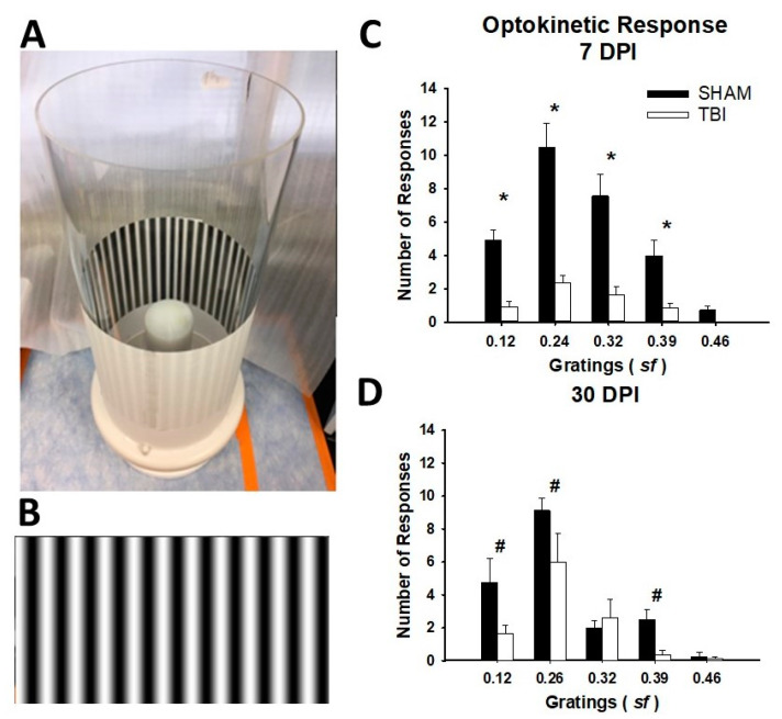
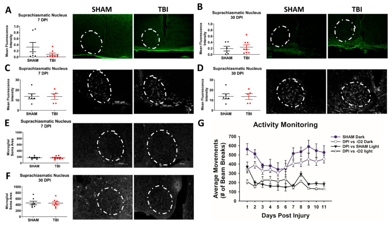
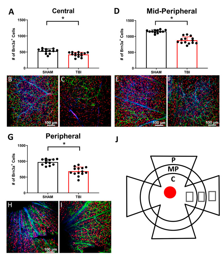
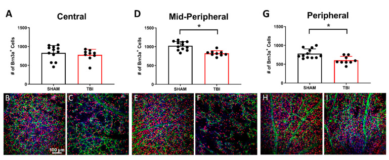
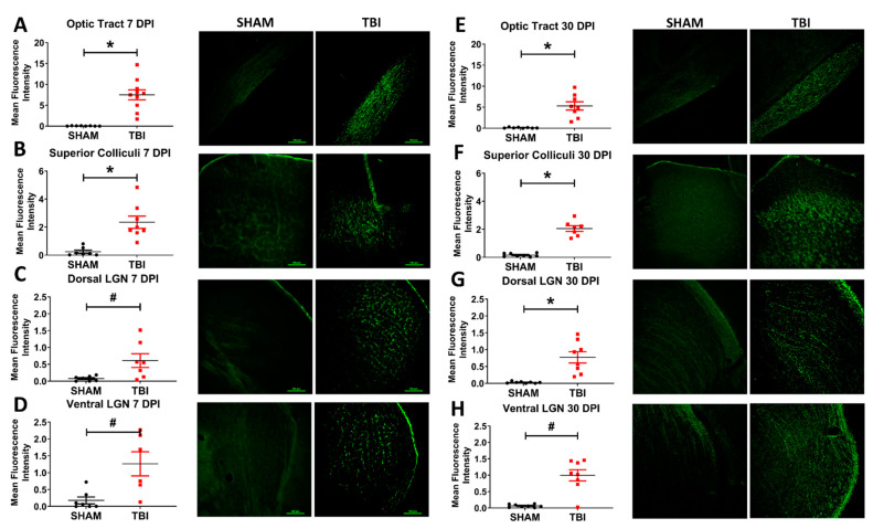
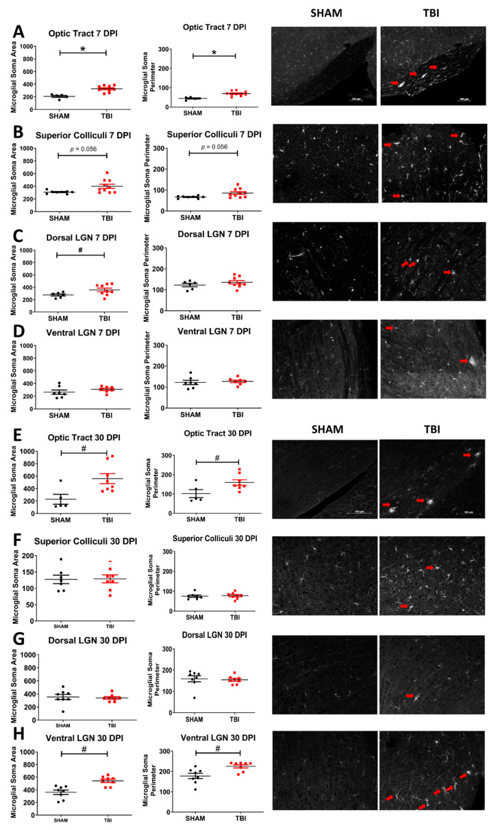
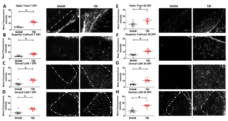
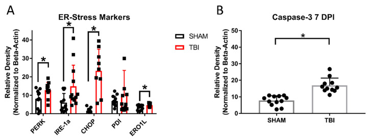
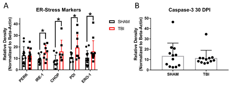
Traumatic brain injury (TBI) results in a number of impairments, often including visual symptoms. In some cases, visual impairments after head trauma are mediated by traumatic injury to the optic nerve, termed traumatic optic neuropathy (TON), which has few effective options for treatment. Using a murine closed-head weight-drop model of head trauma, we previously reported in adult mice that there is relatively selective injury to the optic tract and thalamic/brainstem projections of the visual system. In the current study, we performed blunt head trauma on adolescent C57BL/6 mice and investigated visual impairment in the primary visual system, now including the retina and using behavioral and histologic methods at new time points. After injury, mice displayed evidence of decreased optomotor responses illustrated by decreased optokinetic nystagmus. There did not appear to be a significant change in circadian locomotor behavior patterns, although there was an overall decrease in locomotor behavior in mice with head injury. There was evidence of axonal degeneration of optic nerve fibers with associated retinal ganglion cell death. There was also evidence of astrogliosis and microgliosis in major central targets of optic nerve projections. Further, there was elevated expression of endoplasmic reticulum (ER) stress markers in retinas of injured mice. Visual impairment, histologic markers of gliosis and neurodegeneration, and elevated ER stress marker expression persisted for at least 30 days after injury. The current results extend our previous findings in adult mice into adolescent mice, provide direct evidence of retinal ganglion cell injury after head trauma and suggest that axonal degeneration is associated with elevated ER stress in this model of TON.
Keywords: ER stress; adolescent head trauma; head trauma; mice; traumatic optic neuropathy.
Conflict of interest statement
The authors declare no conflict of interest.
Figures
Similar articles
-
Optic tract injury after closed head traumatic brain injury in mice: A model of indirect traumatic optic neuropathy.PLoS One. 2018 May 10;13(5):e0197346. doi: 10.1371/journal.pone.0197346. eCollection 2018. PLoS One. 2018. PMID: 29746557 Free PMC article.
-
Chronic Histological Outcomes of Indirect Traumatic Optic Neuropathy in Adolescent Mice: Persistent Degeneration and Temporally Regulated Glial Responses.Cells. 2021 Nov 28;10(12):3343. doi: 10.3390/cells10123343. Cells. 2021. PMID: 34943851 Free PMC article.
-
Traumatic Brain Injury-Related Optic Nerve Damage.J Neuropathol Exp Neurol. 2022 Apr 27;81(5):344-355. doi: 10.1093/jnen/nlac018. J Neuropathol Exp Neurol. 2022. PMID: 35363316
-
Connecting endoplasmic reticulum and oxidative stress to retinal degeneration, TBI, and traumatic optic neuropathy.J Neurosci Res. 2020 Mar;98(3):571-574. doi: 10.1002/jnr.24543. Epub 2019 Oct 23. J Neurosci Res. 2020. PMID: 31642095 Review.
-
Afferent Visual Manifestations of Traumatic Brain Injury.J Neurotrauma. 2021 Oct 15;38(20):2778-2789. doi: 10.1089/neu.2021.0182. Epub 2021 Aug 17. J Neurotrauma. 2021. PMID: 34269619 Review.
Cited by
-
Effects of light perception on visual function recovery in patients with traumatic optic neuropathy.Sci Rep. 2024 Mar 29;14(1):7514. doi: 10.1038/s41598-024-54324-1. Sci Rep. 2024. PMID: 38553505 Free PMC article.
-
The Intertwined Roles of Oxidative Stress and Endoplasmic Reticulum Stress in Glaucoma.Antioxidants (Basel). 2022 Apr 29;11(5):886. doi: 10.3390/antiox11050886. Antioxidants (Basel). 2022. PMID: 35624748 Free PMC article. Review.
-
An Update to Biomechanical and Biochemical Principles of Retinal Injury in Child Abuse.Children (Basel). 2024 May 12;11(5):586. doi: 10.3390/children11050586. Children (Basel). 2024. PMID: 38790581 Free PMC article. Review.
-
Neuroprotective Effect of Methylene Blue in a Rat Model of Traumatic Optic Neuropathy.Pharmaceuticals (Basel). 2025 Jun 19;18(6):920. doi: 10.3390/ph18060920. Pharmaceuticals (Basel). 2025. PMID: 40573315 Free PMC article.
-
Complement propagates visual system pathology following traumatic brain injury.J Neuroinflammation. 2024 Apr 17;21(1):98. doi: 10.1186/s12974-024-03098-4. J Neuroinflammation. 2024. PMID: 38632569 Free PMC article.
References
Publication types
MeSH terms
Grants and funding
LinkOut - more resources
Full Text Sources
Medical
