. 2021 Apr 23;11(5):762.
doi: 10.3390/diagnostics11050762.

Evaluation of Correlations between Genetic Variants and High-Resolution Computed Tomography Patterns in Idiopathic Pulmonary Fibrosis

Affiliations

Evaluation of Correlations between Genetic Variants and High-Resolution Computed Tomography Patterns in Idiopathic Pulmonary Fibrosis

Elisa Baratella et al. Diagnostics (Basel). .

Abstract

Background: Idiopathic pulmonary fibrosis (IPF) is a progressive fibrosing interstitial lung disease (ILD). This prospective observational study aimed at the evaluation of any correlation between genetic variants associated with IPF susceptibility and high-resolution computed tomography (HRCT) patterns. It also aimed at evidencing any differences in the HRTC pattern between the familial and sporadic form at diagnosis and after two years.

Methods: A total of 65 IPF patients (mean age at diagnosis 65 ± 10) were enrolled after having given written informed consent. HRCT and genetic evaluations were performed.

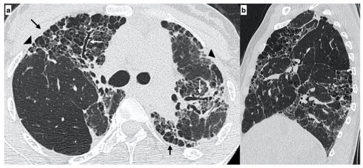
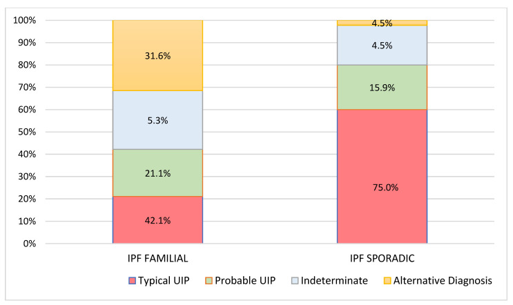
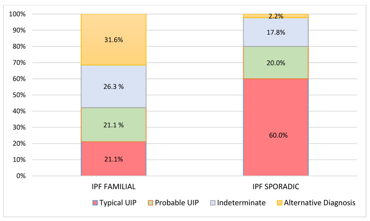
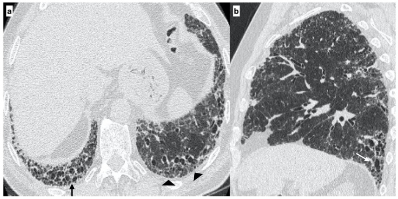
Results: A total of 19 familial (mean age 62 ± 15) and 46 sporadic (mean age 70 ± 9) IPF patients were enrolled. A statistically significant difference was evidenced in the HRTC pattern at diagnosis between the two groups. Sporadic IPF patients had a predominantly usual interstitial pneumonia (UIP) pattern compared with those patients with familial IPF (60.0% vs. 21.1%, respectively). Moreover, familial IPF patients had more alternative diagnoses than those with sporadic IPF (31.6% vs. 2.2%, respectively). Furthermore, there was a slight increase in the typical UIP pattern in the familial IPF group at two years from diagnosis.

Conclusions: Genetic factors play a pivotal role in the risk of developing IPF. However, further studies are required to clarify how these genetic factors may guide clinical treatment decisions.

Keywords: familial idiopathic pulmonary fibrosis; high-resolution computed tomography (HRCT); idiopathic pulmonary fibrosis; interstitial lung disease.


Conflict of interest statement

The authors declare no conflict of interest.

Figures

Figure 1
Figure 1
A 29-year-old female with an “alternative diagnosis pattern” on HRCT at onset of respiratory symptoms. The axial HRCT image shows the presence of honeycombing (black arrows), reticulations (arrowheads) and traction bronchiectasis/bronchiolectasis (white arrows) with a prevalent upper-mid lung distribution (a). The sagittal plane better evidences the atypical distribution of fibrotic alterations (b). Genetic analysis demonstrated the following mutations: ABCA3, SFTPA2, promoter-TERT, SFTPC.
Figure 2
Figure 2
Baseline High-resolution computed tomography (HRCT) patterns with respect to Familial and Sporadic Idiopathic pulmonary fibrosis (IPF).
Figure 3
Figure 3
High-resolution computed tomography (HRCT) patterns at 2 years of follow-up respect to familial and sporadic idiopathic pulmonary fibrosis (IPF).
Figure 4
Figure 4
A 67-year-old male with “UIP pattern” on HRCT at the onset of respiratory symptoms. Axial HRCT plane shows honeycombing (black arrows) and reticulations (arrowheads) with a basal predominant distribution (a); traction bronchiectasis (white arrows) and distribution of alterations are better visible on the sagittal plane (b). Genetic analysis demonstrated the following types of mutations: SFTPA2, promoter-TERT, TERT.
Figure 5
Figure 5
A 64-year-old male with an “Indeterminate pattern” on HRCT at onset of respiratory symptoms. The baseline axial HRCT image evidences subtle ground-glass opacities (black arrows) and reticulation (arrowheads) located in the subpleural regions of the lung bases with a predilection for the peripheral areas of both lungs (a). Follow-up HRCT, performed two years later, evidenced the worsening of the HRCT pattern due to the presence of extensive reticulations (arrowheads) and traction bronchiectasis/bronchiolectasis (white arrows) with a basal predominance (probable UIP pattern) (b). Genetic analysis demonstrated the following mutations: SFTPA2, promoter-TERT, SFTPC.

References

    1. Richeldi L., Collard H.R., Jones M.G. Idiopathic pulmonary fibrosis. Lancet. 2017;389:1941–1952. doi: 10.1016/S0140-6736(17)30866-8. - DOI - PubMed
    1. Hutchinson J.P., Fogarty A.W., Hubbard R.B., McKeever T.M. Global incidence and mortality of idiopathic pulmonary fibrosis: A systematic review. Eur. Respir. J. 2015;46:795–806. doi: 10.1183/09031936.00185114. - DOI - PubMed
    1. Diamantopoulos A., Wright E., Vlahopoulou K., Cornic L., Schoof N., Maher T.M. The Burden of Illness of Idiopathic Pulmonary Fibrosis: A Comprehensive Evidence Review. Pharmacoeconomics. 2018;36:779–807. doi: 10.1007/s40273-018-0631-8. - DOI - PMC - PubMed
    1. Raghu G., Remy-Jardin M., Myers J.L., Richeldi L., Ryerson C.J., Lederer D.J., Behr J., Cottin V., Danoff S.K., Morell F., et al. Diagnosis of Idiopathic Pulmonary Fibrosis. An Official ATS/ERS/JRS/ALAT Clinical Practice Guideline. Am. J. Respir. Crit. Care Med. 2018;198:e44–e68. doi: 10.1164/rccm.201807-1255ST. - DOI - PubMed
    1. Salton F., Ruaro B., Confalonieri P., Confalonieri M. Epithelial–Mesenchymal Transition: A Major Pathogenic Driver in Idiopathic Pulmonary Fibrosis? Medecine. 2020;56:608. doi: 10.3390/medicina56110608. - DOI - PMC - PubMed

LinkOut - more resources