. 2021 Apr 25;13(9):2066.
doi: 10.3390/cancers13092066.

Induction of Apoptosis, Inhibition of MCL-1, and VEGF-A Expression Are Associated with the Anti-Cancer Efficacy of Magnolol Combined with Regorafenib in Hepatocellular Carcinoma

Affiliations

Induction of Apoptosis, Inhibition of MCL-1, and VEGF-A Expression Are Associated with the Anti-Cancer Efficacy of Magnolol Combined with Regorafenib in Hepatocellular Carcinoma

Cheng-Hsien Chen et al. Cancers (Basel). .

Abstract

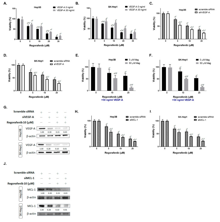
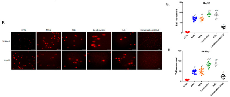
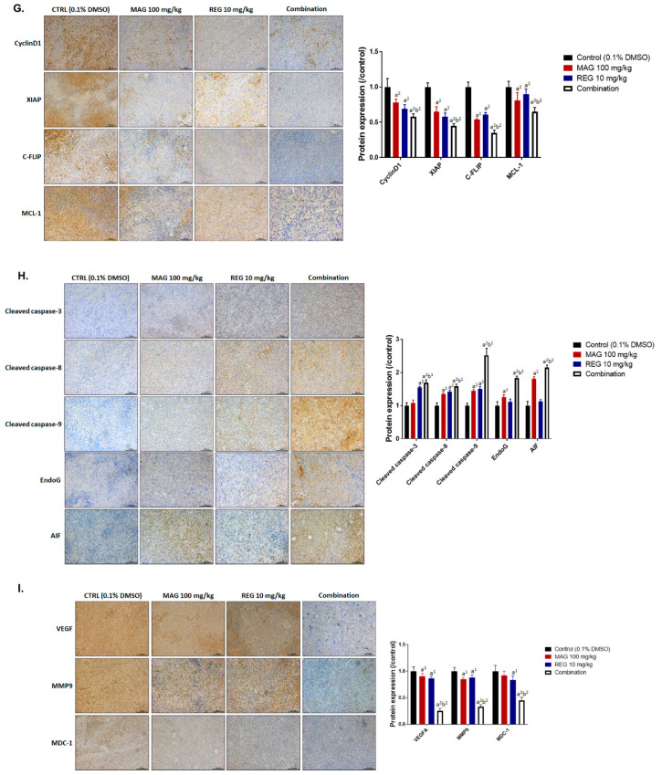
While regorafenib was approved for the treatment of advanced HCC in 2017, with a partial response and survival benefit; other combination agents to facilitate the efficacy of regorafenib still need to be explored. Magnolol is a potential natural anti-tumor compound for many types of cancers. Combination indexes calculated on the basis of both in vitro and in vivo models have indicated a synergistic effect of the combination of regorafenib and magnolol. The overexpression of the VEGF-A protein significantly diminished regorafenib's inhibition of cell viability, while the transient knockdown of VEGF-A by siRNA effectively sensitized HCC cells to regorafenib. In addition, the inhibition of MCL-1 by siRNA combined with regorafenib allowed for a significantly greater inhibition of cell growth, compared to regorafenib alone. A lower protein expression level for VEGF-A and MCL-1 was found for the combination treatment of HCC in vitro and in vivo. A superior metastasis inhibition was also found in the combination group, as compared to the single-treatment groups, using a transwell assay, wound healing assay, and Western blotting. The caspase-dependent and -independent and DNA damage effects, as determined by flow cytometry and a comet assay, were increased by the combination therapy. Taken together, magnolol sensitized HCC to regorafenib, which was correlated with the reduction of VEGF-A and MCL-1 and the induction of apoptosis.

Keywords: MCL-1; VEGF-A; apoptosis; hepatocellular carcinoma; magnolol; regorafenib.


Conflict of interest statement

The authors declare no conflict of interest.

Figures

Figure 1
Figure 1
Cytotoxicity of regorafenib was triggered by magnolol. Hep3B and SK-Hep1 cells were treated with (A) 0–20 μM regorafenib, (B) 0–100 μM magnolol, or (C,D) 50 μM magnolol combined with 0–20 μM regorafenib for 48 h and assayed by the MTT assay. The combination index value for 50 μM magnolol combined with 0–20 μM regorafenib in (E) Hep3B and (F) SK-Hep1 cells is presented. The protein expression of CyclinD1, XIAP, C-FLIP, MCL-1, MMP-9, VEGF-A, and MDC-1 is presented after the treatments with magnolol, regorafenib, and a combination in (G) Hep3B and (H) SK-Hep1 cells. (a1 p < 0.05 and a2 p < 0.01 vs. 0 μM regorafenib or magnolol).
Figure 1
Figure 1
Cytotoxicity of regorafenib was triggered by magnolol. Hep3B and SK-Hep1 cells were treated with (A) 0–20 μM regorafenib, (B) 0–100 μM magnolol, or (C,D) 50 μM magnolol combined with 0–20 μM regorafenib for 48 h and assayed by the MTT assay. The combination index value for 50 μM magnolol combined with 0–20 μM regorafenib in (E) Hep3B and (F) SK-Hep1 cells is presented. The protein expression of CyclinD1, XIAP, C-FLIP, MCL-1, MMP-9, VEGF-A, and MDC-1 is presented after the treatments with magnolol, regorafenib, and a combination in (G) Hep3B and (H) SK-Hep1 cells. (a1 p < 0.05 and a2 p < 0.01 vs. 0 μM regorafenib or magnolol).
Figure 2
Figure 2
Inhibition of VEGF-A and MCL-1 may enhance the cytotoxicity of regorafenib. (A) Hep3B and (B) SK-Hep1 cells were treated with 0–20 μM regorafenib combined with 30 ng/mL of VEGF-A recombinant protein (or not) for 48 h, and the cell viability was measured via the MTT assay. (C) The Hep3B and (D) SK-Hep1 cells were transfected with siVEGF-A combined with 0–20 μM regorafenib (or not) for 48 h, and the cell viability was determined via the MTT assay. (E) The Hep3B and (F) SK-Hep1 cells were treated with 0–20 μM regorafenib, 50 ng/mL of VEGF-A recombinant protein, and 50 μM magnolol for 48 h, and the cell viability was determined via the MTT assay. (G) The VEGF-A Western blotting results in Hep3B and SK-Hep1 cells after being transfected with siVEGF-A combined with 0–20 μM regorafenib (or not) for 48 h. (H) The Hep3B and (I) SK-Hep1 cells were transfected with siMCL-1 combined with 0–20 μM regorafenib (or not) for 48 h, and the cell viability was determined via the MTT assay. (J) The MCL-1 Western blotting results in Hep3B and SK-Hep1 cells after transfection with siMCL-1 combined with 0–20 μM regorafenib (or not) for 48 h. (a1 p < 0.05 and a2 p < 0.01 vs. 0 μM regorafenib; b1 p < 0.05 and b2 p < 0.01 vs. 30 ng/mL VEGF-A; c1 p < 0.05 and c2 p < 0.01 vs. scramble siRNA; d2 p < 0.01 vs. 0 μM MAG).
Figure 3
Figure 3
The metastasis inhibition effect of regorafenib was enhanced by magnolol. Hep3B and SK-Hep1 cells were treated with 10 μM regorafenib, 50 μM magnolol, and a combination for 48 h and subjected to a transwell invasion assay and wound healing assay. (A) A photograph of the invasion transwell and (B) the quantification results of the invasion cells. (C). A photograph of the migration cell pattern and the quantification results of the migrated gap area in (D) the Hep3B and (E) SK-Hep1 cells. (a2 p < 0.01 vs. CTRL; b2 p < 0.01 vs. 10 μM REG or 50 μM MAG; CTRL: Control, MAG: magnolol; REG: regorafenib).
Figure 4
Figure 4
The apoptosis and DNA damage effect of regorafenib was enhanced by magnolol. Hep3B and SK-Hep1 cells were treated with 10 μM regorafenib, 50 μM magnolol, and a combination of both for 48 h, combined with ZVAD for 30 min, and assayed by flow cytometry with (A,B) annexin-V/PI staining and (C) cleaved caspase-3. After 48 h, the magnolol and regorafenib treatments were also performed with (D) PI staining and (E) cleaved PARP-1 staining, respectively. H2O2 was used as a positive control in the comet assay. The tail movement pattern of the Hep3B and SK-Hep1 cells were (F) photographed and (G,H) quantified. (a2 p < 0.01 vs. CTRL; b2 p < 0.01 vs. 10 μM REG or 50 μM MAG; c2 p < 0.01 vs. treatment groups without ZVAD; scale bar = 200 μm).
Figure 4
Figure 4
The apoptosis and DNA damage effect of regorafenib was enhanced by magnolol. Hep3B and SK-Hep1 cells were treated with 10 μM regorafenib, 50 μM magnolol, and a combination of both for 48 h, combined with ZVAD for 30 min, and assayed by flow cytometry with (A,B) annexin-V/PI staining and (C) cleaved caspase-3. After 48 h, the magnolol and regorafenib treatments were also performed with (D) PI staining and (E) cleaved PARP-1 staining, respectively. H2O2 was used as a positive control in the comet assay. The tail movement pattern of the Hep3B and SK-Hep1 cells were (F) photographed and (G,H) quantified. (a2 p < 0.01 vs. CTRL; b2 p < 0.01 vs. 10 μM REG or 50 μM MAG; c2 p < 0.01 vs. treatment groups without ZVAD; scale bar = 200 μm).
Figure 5
Figure 5
The caspase-dependent and -independent apoptotic effects of regorafenib were enhanced by magnolol. Hep3B and SK-Hep1 cells were treated with 10 μM regorafenib, 50 μM magnolol, and a combination for 48 h and assayed by flow cytometry using (A) FAS staining, (B). FAS-L staining, (C) cleaved caspase-8 staining, (D) DCFH-DA staining for ROS, (E) DIOC6 staining for ∆ψm, (F) Fluo-3/AM for Ca2+, and (G) cleaved caspase-9. The Western blotting of BAX, BAK, FAS, FAS-L, cleaved caspase-3 (cleaved C3), -8 (cleaved C8), -9 (cleaved C9), and cleaved PARP-1 in (H) the Hep3B and (I) SK-Hep1 cells. The IF staining of AIF in (J) Hep3B and (K) SK-Hep1 cells. (a2 p < 0.01 vs. CTRL; b2 p < 0.01 vs. 10 μM REG or 50 μM MAG; scale bar = 100 μm).
Figure 5
Figure 5
The caspase-dependent and -independent apoptotic effects of regorafenib were enhanced by magnolol. Hep3B and SK-Hep1 cells were treated with 10 μM regorafenib, 50 μM magnolol, and a combination for 48 h and assayed by flow cytometry using (A) FAS staining, (B). FAS-L staining, (C) cleaved caspase-8 staining, (D) DCFH-DA staining for ROS, (E) DIOC6 staining for ∆ψm, (F) Fluo-3/AM for Ca2+, and (G) cleaved caspase-9. The Western blotting of BAX, BAK, FAS, FAS-L, cleaved caspase-3 (cleaved C3), -8 (cleaved C8), -9 (cleaved C9), and cleaved PARP-1 in (H) the Hep3B and (I) SK-Hep1 cells. The IF staining of AIF in (J) Hep3B and (K) SK-Hep1 cells. (a2 p < 0.01 vs. CTRL; b2 p < 0.01 vs. 10 μM REG or 50 μM MAG; scale bar = 100 μm).
Figure 6
Figure 6
The therapeutic efficacy of regorafenib in Hep3B-bearing mice was enhanced by magnolol. (A) The tumor volume and (B) photographs and (C) weight are displayed after various treatments. (D) Micro-CT scan images, (E) mice body weight, and (F) an H&E stain of the liver, kidneys, and spleen are presented. IHC stain against (G) tumor progression, (H) apoptosis, (I) metastasis- and DNA repair-related antibodies are shown in the tumor tissue of each group. (a1 p < 0.05 and a2 p < 0.01 vs. CTRL; b2 p < 0.01 vs. 10 μM REG or 50 μM MAG; scale bar = 100 μm).
Figure 6
Figure 6
The therapeutic efficacy of regorafenib in Hep3B-bearing mice was enhanced by magnolol. (A) The tumor volume and (B) photographs and (C) weight are displayed after various treatments. (D) Micro-CT scan images, (E) mice body weight, and (F) an H&E stain of the liver, kidneys, and spleen are presented. IHC stain against (G) tumor progression, (H) apoptosis, (I) metastasis- and DNA repair-related antibodies are shown in the tumor tissue of each group. (a1 p < 0.05 and a2 p < 0.01 vs. CTRL; b2 p < 0.01 vs. 10 μM REG or 50 μM MAG; scale bar = 100 μm).

Similar articles

Cited by

References

    1. Grothey A., Blay J.Y., Pavlakis N., Yoshino T., Bruix J. Evolving role of regorafenib for the treatment of advanced cancers. Cancer Treat. Rev. 2020;86:101993. doi: 10.1016/j.ctrv.2020.101993. - DOI - PubMed
    1. D’Alessandro R., Refolo M.G., Iacovazzi P.A., Pesole P.L., Messa C., Carr B.I. Ramucirumab and GSK1838705A Enhance the Inhibitory Effects of Low Concentration Sorafenib and Regorafenib Combination on HCC Cell Growth and Motility. Cancers. 2019;11:787. doi: 10.3390/cancers11060787. - DOI - PMC - PubMed
    1. Mercier J., Voutsadakis I.A. A Systematic Review and Meta-analysis of Retrospective Series of Regorafenib for Treatment of Metastatic Colorectal Cancer. Anticancer Res. 2017;37:5925–5934. doi: 10.21873/anticanres.12039. - DOI - PubMed
    1. Zeiner P.S., Kinzig M., Divé I., Maurer G.D., Filipski K., Harter P.N., Senft C., Bähr O., Hattingen E., Steinbach J.P., et al. Regorafenib CSF Penetration, Efficacy, and MRI Patterns in Recurrent Malignant Glioma Patients. J. Clin Med. 2019;8:2031. doi: 10.3390/jcm8122031. - DOI - PMC - PubMed
    1. Refolo M.G., Lippolis C., Carella N., Cavallini A., Messa C., D’Alessandro R. Chlorogenic Acid Improves the Regorafenib Effects in Human Hepatocellular Carcinoma Cells. Int. J. Mol Sci. 2018;19:1518. doi: 10.3390/ijms19051518. - DOI - PMC - PubMed

LinkOut - more resources