. 2021 Apr 25;10(5):1014.
doi: 10.3390/cells10051014.

Transduction of Pig Small Airway Epithelial Cells and Distal Lung Progenitor Cells by AAV4

Affiliations

Transduction of Pig Small Airway Epithelial Cells and Distal Lung Progenitor Cells by AAV4

Oliver G Chen et al. Cells. .

Abstract

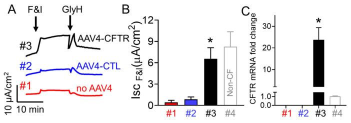
Cystic fibrosis (CF) is caused by genetic mutations of the CF transmembrane conductance regulator (CFTR), leading to disrupted transport of Cl- and bicarbonate and CF lung disease featuring bacterial colonization and chronic infection in conducting airways. CF pigs engineered by mutating CFTR develop lung disease that mimics human CF, and are well-suited for investigating CF lung disease therapeutics. Clinical data suggest small airways play a key role in the early pathogenesis of CF lung disease, but few preclinical studies have focused on small airways. Efficient targeted delivery of CFTR cDNA to small airway epithelium may correct the CFTR defect and prevent lung infections. Adeno-associated virus 4 (AAV4) is a natural AAV serotype and a safe vector with lower immunogenicity than other gene therapy vectors such as adenovirus. Our analysis of AAV natural serotypes using cultured primary pig airway epithelia showed that AAV4 has high tropism for airway epithelia and higher transduction efficiency for small airways compared with large airways. AAV4 mediated the delivery of CFTR, and corrected Cl- transport in cultured primary small airway epithelia from CF pigs. Moreover, AAV4 was superior to all other natural AAV serotypes in transducing ITGα6β4+ pig distal lung progenitor cells. In addition, AAV4 encoding eGFP can infect pig distal lung epithelia in vivo. This study demonstrates AAV4 tropism in small airway progenitor cells, which it efficiently transduces. AAV4 offers a novel tool for mechanistical study of the role of small airway in CF lung pathogenesis in a preclinical large animal model.

Keywords: AAV4; CFTR; cystic fibrosis 1; progenitor cells; small airway epithelia.


Conflict of interest statement

The authors declare no conflict of interest. The funders had no role in the design of the study; in the collection, analyses, or interpretation of data; in the writing of the manuscript, or in the decision to publish the results.

Figures

Figure 1
Figure 1
Identification of integrin (ITG) α6+ cells in the pig distal lung. (A): Pig distal lung was immunostained with Keratin 5 (K5, red), integrin (ITG) α6 (ITGa6, green), and DAPI (blue). Arrow indicates that ITG α6+ progenitor cells (green) are adjacent to K5+ (red) basal cells. (B): FACS assay for immunostained ITGα6+ cells among cultured small airway epithelial cells indicated that 27.5% of the cells are ITGα6+ basal cells. (C): FACS assay for isotype control antibody immunostained cells among cultured small airway epithelial cells. All the cells came from non-CF pigs.
Figure 2
Figure 2
Isolation of ITG α6+/EPCAM+/CD31 progenitor cells in the pig distal lung. The surface expression of ITG α6, EPCAM and CD31 of single cells preparation isolated from a wide type pig lung was analyzed by FACS. (A) SSC vs. FSC density plot. P1 was gated for next step analysis. (B) FSC-Width vs. FSC-Area density plot. P2 was gated to exclude doublets for next step analysis (C) Hoechst 33258 was used to stain dead cells. P3 was gated to select live single cells for next step analysis. (D) CD31+ endothelial cells were excluded by P4 gate using FASC strategy. P5 was gated for next step analysis. Cells positive for both ITG α6 and EPCAM were identified as progenitor cells using P6 gate strategy (E). Black arrows indicate sequential workflow. All the cells came from non-CF pigs.
Figure 3
Figure 3
Screening AAV2H22 and derivatives for tropism for ITG α6β4+ pig progenitor cells. Freshly isolated ITG α6β4+ pig progenitor cells were transduced with the following viral vectors encoding eGFP: AAV2H22 (A), AAV2H22 derivatives viral vectors such as AAV2H22-E67A (B), AAV2H22-S207G (C), AAV2H22-Q598L (D), AAV2H22-I648V (E), AAV2H22-V708I (F). GFP+ cluster were quantified after progenitor cells were cultured in the Matrigel for 2 weeks. The percentage of GFP+ colonies among the entire progenitor cells for each viral vector were quantified (G). All the cells came from non-CF pigs. Scale bars in all images = 1 mm.
Figure 4
Figure 4
Screening AAV2 and derivatives for tropism for α6β4+ pig progenitor cells. Freshly isolated ITG α6β4+ pig progenitor cells were transduced with the following viral vectors encoding eGFP: AAV2 (A), AAV2 derivatives viral vectors including AAV2-E67A (B), AAV2-S207G (C), AAV2-Q598L (D), AAV2-I648V (E). GFP+ clusters were quantified after progenitor cells were cultured in the Matrigel for 2 weeks. Quantification of the percentage of GFP+ colonies among the entire progenitor cells for each viral vector shows maximum transduction efficiency was less than 15% (F). All the cells came from non-CF pigs. Scale bars in all images = 1 mm.
Figure 5
Figure 5
Screening AAV natural serotypes for tropism for α6β4+ pig progenitor cells and AAV4 was far superior to all other serotypes. Freshly isolated ITG α6β4+ pig progenitor cells were transduced with the following viral vectors encoding eGFP: AAV1 (A), AAV2 (B), AAV4 (C), AAV5 (D), AAV6 (E), AAV8 (F), AAV9 (G), AAV-DJ (H). GFP+ clusters were quantified after progenitor cells were cultured in the Matrigel for 2 weeks. Quantification of the percentage of GFP+ colonies among the entire progenitor cells for each viral vector shows that AAVV4 has the highest transduction efficiency (I). All the cells came from non-CF pigs. Scale bars in all images = 1 mm.
Figure 6
Figure 6
AAV4 transduce both large and small airway epithelial cells but with much higher tropism for small airways. AAV4 encoding eGFP transduces both large (A) and small (B) pig airway epithelial cells. Scale bars = 500µm. (C) Quantification of AAV4-GFP transduced GFP+ clusters demonstrate much higher expression in epithelial cells from pig small airways compared with large airways. N = 5, * p < 0.05 compared to large airways cells. All the cells came from non-CF pigs. Scale bars in all images = 1 mm.
Figure 7
Figure 7
Lectin binding profiles assays in large and small porcine airway epithelium. There is much higher expression of O-linked carbohydrates and O-glycoproteins demonstrated by fluorescence assay for jacalin (A,B) in large airways compared with small airways. In contrast, there is much higher expression of N-acetyl-D-glucosamine demonstrated by wheat germ agglutinin assay (WGA, (C,D)) in small airways compared to large airways. In addition, there is more 2,3-linked sialic acid demonstrated by MAA (E,F) fluorescence assay in large airways compared to small airways. Moreover, there is more mannose demonstrated by ConA (G,H) fluorescence assay in small airways compared to large airways. All the cells came from non-CF pigs.
Figure 8
Figure 8
AAV4 encoding CFTR significantly increased cAMP stimulated short circuit current (Isc) and CFTR gene expression in CF pig small airway epithelia. (A) Sample Ussing chamber traces from small airway epithelial cells from CF pigs in response to indicated chemical treatments. Trace #1: control without AAV4 treatment; #2: CF cells treated with AAV4 encoding GFP; #3: CF cells treated with AAV4 encoding CFTR. (B) Summary of cAMP stimulated Isc. N = 3, * p < 0.05 compared to #1 and #2. #4, non-CF cells. (C) AAV4 encoding CFTR increased CFTR expression in CF cells. N = 3. * p < 0.05 compared to #1 and #2. #4, non-CF cells.
Figure 9
Figure 9
AAV4-mediated transgene expression in pig distal lung. AAV4-CMV > eGFP was administered via bronchoscope to pig distal lung. Immunofluorescent imaging of tissue confirmed expression of GFP+ cells in bronchioalveolar junction (A), alveolar region (B), and small airways (C) in vivo. All the images came from non-CF pigs. N = 3.

References

    1. Welsh M.J., Smith A.E. Cystic fibrosis. Sci. Am. 1995;273:52–59. doi: 10.1038/scientificamerican1295-52. - DOI - PubMed
    1. Stoltz D.A., Meyerholz D.K., Welsh M.J. Origins of cystic fibrosis lung disease. N. Engl. J. Med. 2015;372:351–362. doi: 10.1056/NEJMra1300109. - DOI - PMC - PubMed
    1. Wainwright C.E., Elborn J.S., Ramsey B.W., Marigowda G., Huang X., Cipolli M., Colombo C., Davies J.C., De Boeck K., Flume P.A., et al. Lumacaftor-ivacaftor in patients with cystic fibrosis homozygous for Phe508del CFTR. N. Engl. J. Med. 2015;373:220–231. doi: 10.1056/NEJMoa1409547. - DOI - PMC - PubMed
    1. Stoltz D.A., Meyerholz D.K., Pezzulo A.A., Ramachandran S., Rogan M.P., Davis G.J., Hanfland R.A., Wohlford-Lenane C., Dohrn C.L., Bartlett J.A., et al. Cystic fibrosis pigs develop lung disease and exhibit defective bacterial eradication at birth. Sci. Transl. Med. 2010;2:29ra31. doi: 10.1126/scitranslmed.3000928. - DOI - PMC - PubMed
    1. Steines B., Dickey D.D., Bergen J., Excoffon K.J.D.A., Weistein J.R., Li X., Yan Z., Alaiwa M.H.A., Shah V.S., Bouzek D.C., et al. CFTR gene transfer with AAV improves early cystic fibrosis pig phenotypes. JCI Insight. 2016;1:e88728. doi: 10.1172/jci.insight.88728. - DOI - PMC - PubMed

Publication types

MeSH terms

Substances