

Ceramic Biomaterial Pores Stereology Analysis by the Use of Microtomography
- PMID: 33923089
- PMCID: PMC8123274
- DOI: 10.3390/ma14092207
Ceramic Biomaterial Pores Stereology Analysis by the Use of Microtomography
Abstract
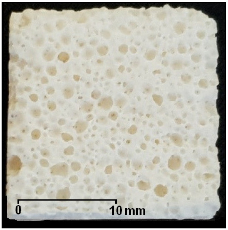
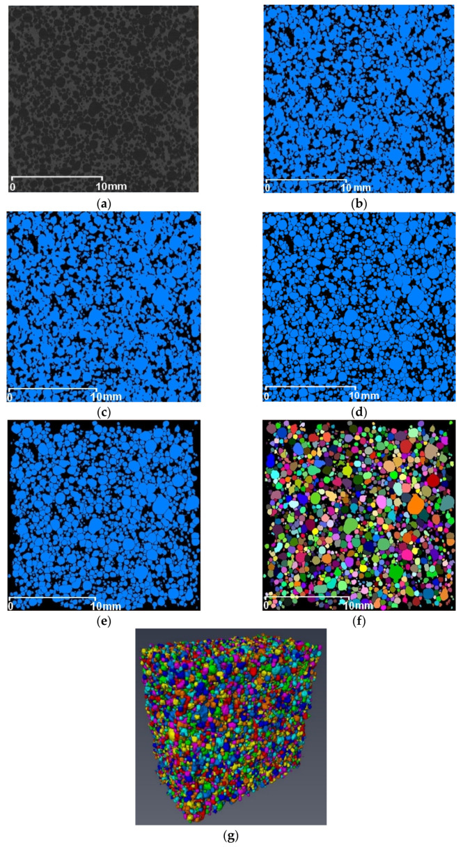
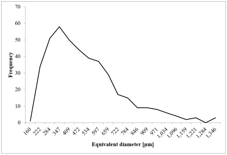
The main aim of this study was to analyze microtomographic data to determine the geometric dimensions of a ceramic porous material's internal structure. Samples of a porous corundum biomaterial were the research material. The samples were prepared by chemical foaming and were measured using an X-ray scanner. In the next stage, 3D images of the samples were generated and analyzed using Thermo Scientific Avizo software. The analysis enabled the isolation of individual pores. Then, the parameters characterizing the pore geometry and the porosity of the samples were calculated. The last part of the research consisted of verifying the developed method by comparing the obtained results with the parameters obtained from the microscopic examinations of the biomaterial. The comparison of the results confirmed the correctness of the developed method. The developed methodology can be used to analyze biomaterial samples to assess the geometric dimensions of biomaterial pores.
Keywords: alumina; image processing; microtomography apparatus; porous bioceramics.
Conflict of interest statement
The authors declare no conflict of interest.
Figures


Similar articles
-
Quantitative stereological analysis of the highly porous hydroxyapatite scaffolds using X-ray CM and SEM.Biomed Mater Eng. 2017;28(3):235-246. doi: 10.3233/BME-171670. Biomed Mater Eng. 2017. PMID: 28527187
-
Pore-throat structure characterization of carbon fiber reinforced resin matrix composites: Employing Micro-CT and Avizo technique.PLoS One. 2021 Sep 22;16(9):e0257640. doi: 10.1371/journal.pone.0257640. eCollection 2021. PLoS One. 2021. PMID: 34551013 Free PMC article.
-
Polyurethane-Based Porous Carbons Suitable for Medical Application.Materials (Basel). 2022 May 5;15(9):3313. doi: 10.3390/ma15093313. Materials (Basel). 2022. PMID: 35591653 Free PMC article.
-
Micro-computed tomographic and SEM study of porous bioceramics using an adaptive method based on the mathematical morphological operations.Heliyon. 2019 Dec 19;5(12):e02557. doi: 10.1016/j.heliyon.2019.e02557. eCollection 2019 Dec. Heliyon. 2019. PMID: 31890929 Free PMC article. Review.
-
3D printed porous ceramic scaffolds for bone tissue engineering: a review.Biomater Sci. 2017 Aug 22;5(9):1690-1698. doi: 10.1039/c7bm00315c. Biomater Sci. 2017. PMID: 28686244 Review.
References
-
- Mour M., Das D., Winkler T., Hoenig E., Mielke G., Morlock M.M., Schilling A.F. Advances in Porous Biomaterials for Dental and Orthopaedic Applications. Materials. 2010;3:2947–2974. doi: 10.3390/ma3052947. - DOI
-
- Szczepkowska M., Łuczuk M. Porous materials for the medical applications. Syst. Wspomagania Inżynierii Prod. 2014;2:231–239.
-
- Paciorek J. Materiały porowate—Właściwości i zastosowanie. Trendy ve vzdělávání. 2012;5:229–232.
-
- Živić F., Grujović N., Mitrović S., Adamović D., Petrović V., Radovanović A., Durić S., Palića N. Friction and Adhesion in Porous Biomaterial Structure. Tribol. Ind. 2016;38:361–370.
Grants and funding
LinkOut - more resources
Full Text Sources
Other Literature Sources
