Review
. 2021 Apr 24;10(5):661.
doi: 10.3390/antiox10050661.

Glutathione Metabolism and the Novel Role of Mitochondrial GSH in Retinal Degeneration

Affiliations
Review

Glutathione Metabolism and the Novel Role of Mitochondrial GSH in Retinal Degeneration

Parameswaran G Sreekumar et al. Antioxidants (Basel). .

Abstract

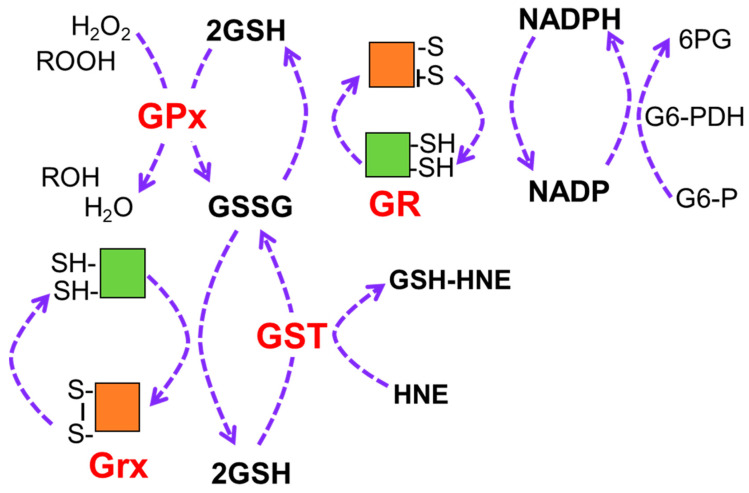
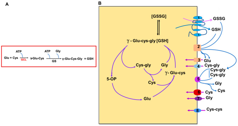
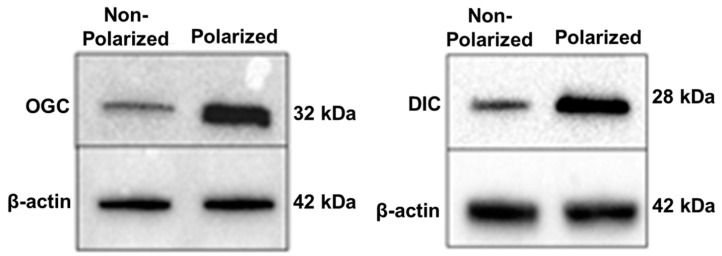
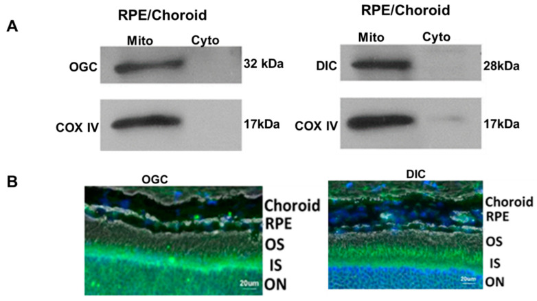
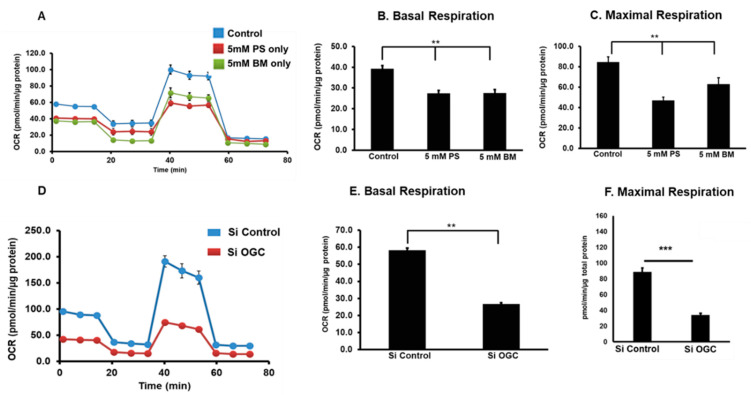
Glutathione (GSH) is present ubiquitously, and its role as a crucial cellular antioxidant in tissues, including the retina, is well established. GSH's antioxidant function arises from its ability to scavenge reactive oxygen species or to serve as an essential cofactor for GSH S-transferases and peroxidases. This review summarizes the general functions, retinal distribution, disorders linked to GSH deficiency, and the emerging role for mitochondrial GSH (mGSH) in retinal function. Though synthesized only in the cytosol, the presence of GSH in multiple cell organelles suggests the requirement for its active transport across organellar membranes. The localization and distribution of 2-oxoglutarate carrier (OGC) and dicarboxylate carrier (DIC), two recently characterized mitochondrial carrier proteins in RPE and retina, show that these transporters are highly expressed in human retinal pigment epithelium (RPE) cells and retinal layers, and their expression increases with RPE polarity in cultured cells. Depletion of mGSH levels via inhibition of the two transporters resulted in reduced mitochondrial bioenergetic parameters (basal respiration, ATP production, maximal respiration, and spare respiratory capacity) and increased RPE cell death. These results begin to reveal a critical role for mGSH in maintaining RPE bioenergetics and cell health. Thus, augmentation of mGSH pool under GSH-deficient conditions may be a valuable tool in treating retinal disorders, such as age-related macular degeneration and optic neuropathies, whose pathologies have been associated with mitochondrial dysfunction.

Keywords: RPE; SLC25A10 (DIC); SLC25A11 (OGC); bioenergetics; mitochondrial GSH; retinal degeneration.


Conflict of interest statement

D.A.F. is a member of the Scientific Advisor Board for Vinci Pharmaceuticals, Inc. R.K. and P.G.S. declare no conflict of interest. The funders had no role in the design of the study; in the collection, analyses, or interpretation of data; in the writing of the manuscript, or in the decision to publish the results.

Figures

Figure 1
Figure 1
Redox cycling regenerates antioxidants and requires GSH. Antioxidants undergo multiple rounds of reduction–oxidation at active site cysteine residues, which are represented by the disulfide (S-S) and SH, respectively. GSH provides the reducing equivalents for these reactions involving the reduction in harmful hydrogen peroxide and organic peroxides (ROOH) by GPx or neutralization of reactive aldehydes, such as HNE, through addition of GSH by GST. GSSG is replenished to GSH by either NADP-dependent GR or Grx. NADPH is produced by reaction with glucose 6-phosphate dehydrogenase. NADPH, nicotinamide adenine dinucleotide phosphate; GSH, reduced glutathione; GSSR, oxidized glutathione; GR, glutathione reductase; GPx, glutathione peroxidase; Grx, glutaredoxin; GST, glutathione S-transferase; G6-PDH, glucose 6-phosphate dehydrogenase.
Figure 2
Figure 2
Scheme showing the biosynthesis of GSH (A), and the glutathione cycle and transport of constituent amino acids and related substrates (B). Figure 2B is modified from Bachhawat AK, Yadav S. The glutathione cycle: glutathione metabolism beyond the γ-glutamyl cycle. IUBMB Life. 2018 July; 70(7):585–592. doi: 10.1002/iub.1756. Epub 2018 Apr 17. PMID: 29667297. Wiley Publishers [19]. GCL—glutamate–cysteine ligase, GS—glutathione synthetase, GSH—glutathione. 5-OP-5-oxoproline. Numbers refer to representative transporters involved. 1–2. MRP family; 3. EAATS family; 4. Peptide transporters; 5–6. System Xc and other transporters; 7. Glycine transporter; 8. Disulfide transporter.
Figure 3
Figure 3
Effect of oxidative stress on cell survival (A) and GSH levels (B) in RPE isolated from AMD donors and age-matched controls (no AMD). Results were measured 24 h after RPEs were exposed to different doses of hydrogen peroxide. Modified from Redox Biol. 2017; 13:255–265, Ferrington et al. [21]. Copyright (2021), with permission obtained from Elsevier. * p < 0.05 as determined by 2-way ANOVA and Tukey’s post-hoc test. Results from 2-way ANOVA for disease (Dis), peroxide dose (Per), and their interaction (DxP) are shown on the graphs.
Figure 4
Figure 4
Polarity upregulates mitochondrial carrier proteins, OGC and DIC, in primary human RPE cells. Polarized RPE cultures had an average TER of 380 ± 60 Ω·cm2 (reproduced from [97] and is licensed under a Creative Commons Attribution-NonCommercial-NoDerivatives 4.0 International License).
Figure 5
Figure 5
Expression of mGSH carrier proteins in mouse RPE/Choroid (A) and whole retina (B). Mitochondria was isolated as described earlier [97], and the specificity of expression in mitochondria is illustrated with COX IV as a mitochondria specific marker in A. Immunofluorescence staining of OGC (green) and DIC (green) in retinal layers is shown in B. Mito: mitochondria, Cyto: cytosol, COX IV: cytochrome c oxidase subunit 4, Reproduced from [97] and is licensed under a Creative Commons Attribution-NonCommercial-NoDerivatives 4.0 International License. Blue: nuclear stain, DAPI. OS—outer segment, IS—inner segment, ON—outer nuclear layer.
Figure 6
Figure 6
Chemical inhibition of OGC and DIC with PS and BM decreased mitochondrial bioenergetics in RPE (AC). Silencing OGC caused a significant decrease in respiratory parameters (DF). (Modified from Sreekumar et al. [81] and is licensed under a Creative Commons Attribution-NonCommercial-NoDerivatives 4.0 International License). ** p < 0.1 *** p < 0.01.

References

    1. Sreekumar P.G., Spee C., Ryan S.J., Cole S.P.C., Kannan R., Hinton D.R. Mechanism of RPE Cell Death in α-Crystallin Deficient Mice: A Novel and Critical Role for MRP1-Mediated GSH Efflux. PLoS ONE. 2012;7:e33420. doi: 10.1371/journal.pone.0033420. - DOI - PMC - PubMed
    1. Kwon D.H., Lee H., Park C., Hong S.-H., Hong S.H., Kim G.-Y., Cha H.-J., Kim S., Kim H.-S., Hwang H.-J., et al. Glutathione Induced Immune-Stimulatory Activity by Promoting M1-Like Macrophages Polarization via Potential ROS Scavenging Capacity. Antioxidants. 2019;8:413. doi: 10.3390/antiox8090413. - DOI - PMC - PubMed
    1. Sinha R., Sinha I., Calcagnotto A., Trushin N., Haley J.S., Schell T.D., Richie J.P. Oral supplementation with liposomal glutathione elevates body stores of glutathione and markers of immune function. Eur. J. Clin. Nutr. 2018;72:105–111. doi: 10.1038/ejcn.2017.132. - DOI - PMC - PubMed
    1. Liu R.M. Gaston Pravia KA: Oxidative stress and glutathione in TGF-beta-mediated fibrogenesis. Free Radic. Biol. Med. 2010;48:1–15. doi: 10.1016/j.freeradbiomed.2009.09.026. - DOI - PMC - PubMed
    1. Ballatori N., Krance S.M., Marchan R., Hammond C.L. Plasma membrane glutathione transporters and their roles in cell physiology and pathophysiology. Mol. Asp. Med. 2009;30:13–28. doi: 10.1016/j.mam.2008.08.004. - DOI - PMC - PubMed

LinkOut - more resources