Melanin Transfer in the Epidermis: The Pursuit of Skin Pigmentation Control Mechanisms
- PMID: 33923362
- PMCID: PMC8123122
- DOI: 10.3390/ijms22094466
Melanin Transfer in the Epidermis: The Pursuit of Skin Pigmentation Control Mechanisms
Abstract
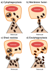
The mechanisms by which the pigment melanin is transferred from melanocytes and processed within keratinocytes to achieve skin pigmentation remain ill-characterized. Nevertheless, several models have emerged in the past decades to explain the transfer process. Here, we review the proposed models for melanin transfer in the skin epidermis, the available evidence supporting each one, and the recent observations in favor of the exo/phagocytosis and shed vesicles models. In order to reconcile the transfer models, we propose that different mechanisms could co-exist to sustain skin pigmentation under different conditions. We also discuss the limited knowledge about melanin processing within keratinocytes. Finally, we pinpoint new questions that ought to be addressed to solve the long-lasting quest for the understanding of how basal skin pigmentation is controlled. This knowledge will allow the emergence of new strategies to treat pigmentary disorders that cause a significant socio-economic burden to patients and healthcare systems worldwide and could also have relevant cosmetic applications.
Keywords: keratinocyte; melanin; melanocore; melanocyte; melanokerasome; melanosome; skin pigmentation.
Conflict of interest statement
The authors declare no conflict of interest.
Figures


References
-
- Lai-Cheong J.E., McGrath J.A. Structure and Function of Skin, Hair and Nails. Medicine. 2013;41:317–320. doi: 10.1016/j.mpmed.2013.04.017. - DOI
-
- Cichorek M., Wachulska M., Stasiewicz A. Heterogeneity of Neural Crest-Derived Melanocytes. Cent. Eur. J. Biol. 2013;8:315–330. doi: 10.2478/s11535-013-0141-1. - DOI
-
- Lapedriza A., Petratou K., Kelsh R.N. Neural Crest Cells and Pigmentation. Neural Crest Cells Evol. Dev. Dis. 2014:287–311. doi: 10.1016/B978-0-12-401730-6.00015-6. - DOI
Publication types
MeSH terms
Substances
Grants and funding
LinkOut - more resources
Full Text Sources
Miscellaneous

