. 2021 Apr 16;10(4):337.
doi: 10.3390/biology10040337.

Biochemical Characterization of the Amylase Activity from the New Haloarchaeal Strain Haloarcula sp. HS Isolated in the Odiel Marshlands

Affiliations

Biochemical Characterization of the Amylase Activity from the New Haloarchaeal Strain Haloarcula sp. HS Isolated in the Odiel Marshlands

Patricia Gómez-Villegas et al. Biology (Basel). .

Abstract

Alpha-amylases are a large family of α,1-4-endo-glycosyl hydrolases distributed in all kingdoms of life. The need for poly-extremotolerant amylases encouraged their search in extreme environments, where archaea become ideal candidates to provide new enzymes that are able to work in the harsh conditions demanded in many industrial applications. In this study, a collection of haloarchaea isolated from Odiel saltern ponds in the southwest of Spain was screened for their amylase activity. The strain that exhibited the highest activity was selected and identified as Haloarcula sp. HS. We demonstrated the existence in both, cellular and extracellular extracts of the new strain, of functional α-amylase activities, which showed to be moderately thermotolerant (optimum around 60 °C), extremely halotolerant (optimum over 25% NaCl), and calcium-dependent. The tryptic digestion followed by HPLC-MS/MS analysis of the partially purified cellular and extracellular extracts allowed to identify the sequence of three alpha-amylases, which despite sharing a low sequence identity, exhibited high three-dimensional structure homology, conserving the typical domains and most of the key consensus residues of α-amylases. Moreover, we proved the potential of the extracellular α-amylase from Haloarcula sp. HS to treat bakery wastes under high salinity conditions.

Keywords: amylase; enzymatic characterization; extremozymes; haloarchaea; proteomics.


Conflict of interest statement

The authors declare no conflict of interest. The funders had no role in the design of the study; in the collection, analyses, or interpretation of data; in the writing of the manuscript, or in the decision to publish the results.

Figures

Figure 1
Figure 1
(A) In vitro selective screening for amylase-releasing haloarchaea. Semi-quantitative estimation of amylase activity from four haloarchaeal strains isolated from the Odiel Marshlands, grown on starch agar plates (top) and revealed with Lugol´s iodine solution (down), as detailed in the Material and Methods section. (B) Molecular phylogenetic analysis by the maximum likelihood method. The tree represents a comparison among the complete 16S rRNA coding gene sequences, including a series of reference haloarchaeal species and the new isolated strain, Haloarcula sp. HS. Multiple alignments were generated by MUSCLE (MUltiple Sequence Comparison by Log-Expectation) and the tree was constructed with MEGA X. The numbers at the nodes indicate the bootstrap values calculated for 1000 replicates. Arrows point to the new strain Haloarcula sp. HS.
Figure 2
Figure 2
Time course evolution of Haloarcula sp. HS cultures in rich and minimal media. Optical density (A), secretion of proteins (B), and starch hydrolysis (C) were measured during the time of culture in rich (■) and minimal () broths. All data are expressed as the mean ± SD of at least triplicate experiments.
Figure 3
Figure 3
Time course evolution of Haloarcula sp. HS in the two-step culture. Optical density, secreted proteins, and starch concentration were measured along the full cultivation time. The red arrow indicates the moment of transference of the cells from the rich to the fresh minimum medium. All data are expressed as the mean ± SD of at least triplicate experiments.
Figure 4
Figure 4
Native-PAGE and zymogram of cell-associated (A) and extracellular (B) proteins obtained from Haloarcula sp. HS. Lanes 1, 3, and 5—samples on native-PAGE followed by Coomassie Blue staining. Lanes 2, 4, and 6—samples on native PAGE followed by Lugol´s solution staining. Lane M—molecular mass marker in kDa, lanes 1 and 2—crude extract, lanes 3 and 4—partially purified crude extract, and lanes 5 and 6—concentrated supernatant. The whole gels for each staining are available in Supplementary Material, Figure S2.
Figure 5
Figure 5
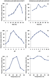
Effect of salt, pH, and temperature on amylase activities. Relative amylase activities in different salt concentrations (%, w/v), pH values, and temperatures are shown for the extracellular (A,C,E) and cellular (B,D,F) extracts. Relative activity was defined as the percentage of maximum activity for each case. The 100% activity corresponded to 70 ± 6.4 U mL−1 (350 U mg−1) for the extracellular amylase extract and 60 ± 5.6 U mL−1 (120 U mg−1) for the mix of cell-associated amylases. Mean and standard deviations are shown.
Figure 6
Figure 6
Effect of metal ions on the amylase activities. Extracellular and cell-associated relative amylase activities under the presence of different metal ions and EDTA (10 mM) are represented. Relative activity was defined as the percentage of maximal activity with respect to control, with no additives. The control activity was 70 ± 6.4 U mL−1 (350 U mg−1) for the extracellular amylase and 60 ± 5.6 U mL−1 (120 U mg−1) for the cellular amylase. Mean and standard deviations are shown.
Figure 7
Figure 7
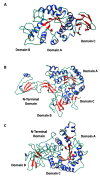
Three-dimensional predicted structures of the three amylase sequences identified in Haloarcula sp. HS; (A) mature extracellular amylase, AMY_HS1; (B) cellular amylase, AMY_HS2; (C) cellular amylase, AMY_HS3. Phyre2 software and the Chimera program were employed for 3D structure visualization. Helixes are represented by blue ribbons and strands by red arrows.
Figure 8
Figure 8
Alignment of the three amylase sequences from Haloarcula sp. HS. The mature extracellular protein is named AMY_HS1, while the cell-associated amylases are denoted as AMY_HS2 and AMY_HS3. Purple stars highlight the catalytic triad (Asp-Glu-Asp), blue star denotes the canonical calcium-binding site and the black stars point other essential residues for enzyme structure. Identical residues in the three sequences are shaded in white, residues that coincide in two of the sequences or do not coincide at all are shaded in pink and red, respectively.

Similar articles

Cited by

References

    1. Kanekar P.P., Kelkar A.S., Dhakephalkar P.K. Halophiles—Taxonomy, Diversity, Physiology and Applications. In: Metzler J.B., editor. Microorganisms in Environmental Management. Volume 9789400722. Springer; Dordrecht, The Netherlands: 2012. pp. 1–34.
    1. Ventosa A., Márquez M.C., Sánchez-Porro C., De La Haba R.R. Taxonomy of Halophilic Archaea and Bacteria. In: Vreeland R.H., editor. Advances in Understanding the Biology of Halophilic Microorganisms. Springer; Dordrecht, The Netherlands: 2012. pp. 59–80.
    1. Oren A. Life at high salt concentrations, intracellular KCl concentrations, and acidic proteomes. Front. Microbiol. 2013;4:315. doi: 10.3389/fmicb.2013.00315. - DOI - PMC - PubMed
    1. Mevarech M., Frolow F., Gloss L.M. Halophilic enzymes: Proteins with a grain of salt. Biophys. Chem. 2000;86:155–164. doi: 10.1016/S0301-4622(00)00126-5. - DOI - PubMed
    1. Mehta D., Satyanarayana T. Bacterial and Archaeal α-Amylases: Diversity and Amelioration of the Desirable Characteristics for Industrial Applications. Front. Microbiol. 2016;7:1129. doi: 10.3389/fmicb.2016.01129. - DOI - PMC - PubMed