Review
. 2021 Apr 16;22(8):4134.
doi: 10.3390/ijms22084134.

The Multifaceted Roles of Ku70/80

Affiliations
Review

The Multifaceted Roles of Ku70/80

Sayma Zahid et al. Int J Mol Sci. .

Abstract

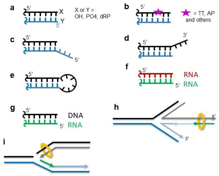
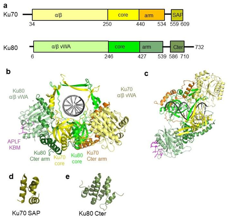
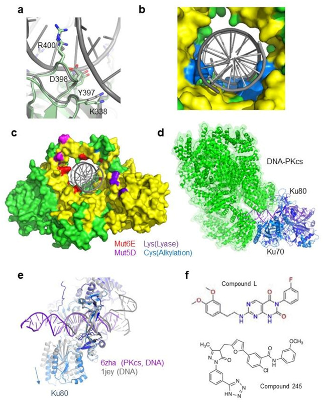
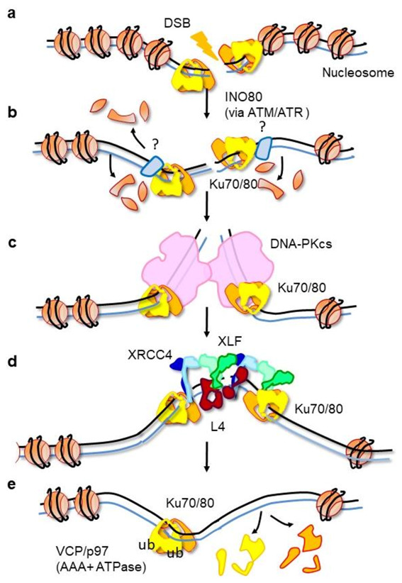
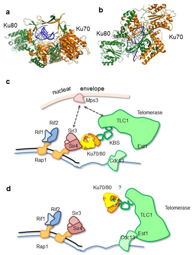
DNA double-strand breaks (DSBs) are accidental lesions generated by various endogenous or exogenous stresses. DSBs are also genetically programmed events during the V(D)J recombination process, meiosis, or other genome rearrangements, and they are intentionally generated to kill cancer during chemo- and radiotherapy. Most DSBs are processed in mammalian cells by the classical nonhomologous end-joining (c-NHEJ) pathway. Understanding the molecular basis of c-NHEJ has major outcomes in several fields, including radiobiology, cancer therapy, immune disease, and genome editing. The heterodimer Ku70/80 (Ku) is a central actor of the c-NHEJ as it rapidly recognizes broken DNA ends in the cell and protects them from nuclease activity. It subsequently recruits many c-NHEJ effectors, including nucleases, polymerases, and the DNA ligase 4 complex. Beyond its DNA repair function, Ku is also involved in several other DNA metabolism processes. Here, we review the structural and functional data on the DNA and RNA recognition properties of Ku implicated in DNA repair and in telomeres maintenance.

Keywords: DNA repair machinery; c-NHEJ; double-strand break; protein-DNA interactions; telomeres.


Conflict of interest statement

The authors declare no conflict of interest.

Figures

Figure 1
Figure 1
Diversity of DNA ends that are recognized by Ku70/80. (a) Ku binds DNA ends efficiently with strands containing either hydroxyl or phosphate functions, as well as more heterogeneous ends with dRP or other chemical adducts. (b) Ku recognizes DSBs with a lesion present in the internal position close to the DNA ends. (c,d) Ku interacts efficiently with DNA having single-strand overhangs in 3′ and/or in 5′ up to 20 nt. (e) Ku can accommodate the presence of loops up to at least 8 nt at the ends. (f) Ku binds RNA hairpins at telomeres. (g,h) Ku interacts with the DNA–RNA duplex during replication fork restart (nascent RNA (green arrow)). (i) Ku binds single-ended DSB.
Figure 2
Figure 2
Structure of the human Ku70/80 heterodimer. (a) Human Ku70 and Ku80 have similar organizations. They present some sequence variabilities in their C-terminal region. (b,c) Crystal structure of Ku70/80 complexed with a hairpin DNA and the Ku binding motif of APLF (magenta) [26]. Ku is colored in the same way as that in (a). The right view shows the top view of the complex. The DNA is a hairpin DNA used to limit Ku movement on the DNA for crystallization. (d) Structure of the SAP domain of Ku70 solved by NMR [48]. (e) C-terminal domain of Ku80 solved by NMR [49,50].
Figure 3
Figure 3
Complementarity between Ku inner ring and the DNA (a) Structure of an inner ring loop of Ku (Ku80 Tyr397-Arg400) that interacts with the minor groove of the DNA. The figure shows a superimposition of this loop in the Ku/DNA complex and Ku free structures. (b) Surface representation of the Ku inner ring. Ku residues in contact with the DNA grooves are colored in blue. Due to these contacts, the radius of the inner ring of Ku is smaller than the DNA radius. (c) Surface representation of Ku. Residues in red are the six Ku70 positions mutated in Glu to disrupt the Ku-DNA interaction [52] (positions Lys282, Lys287, Thr300, Lys331, Lys338, and Arg403). Residues in magenta are the five positions mutated in Asp in [53] to mimic potential phosphorylation sites on the ring (Thr305, Ser306, Thr307, Ser314, and Thr316). Residues in purple are positions of Ku70 lysines (Lys31, Lys160, and Lys164) proposed to be involved in the Ku dRP/Lyase activity [43] (Lys31 is not observe, the first amino acid of Ku70 visible in the crystal is Arg35). Residues in light blue are a Cys of Ku80 that is a potential site of alkylation [54]. (d) CryoEM structure of (Ku70/80)/DNA-PKcs/DNA complex (PDB 6zha) [11]. DNA-PKcs is represented in the green cartoon with a semi-transparent surface. Ku70 and Ku80 are, respectively, shown in light and dark blue, while DNA is shown in purple. (e) Superimposition of Ku from the Ku/DNA-PKcs/DNA complex and Ku from the Ku/DNA complex (PDB 1jey) [45]. The loops of Ku in contact with DNA superimpose well, indicating that Ku has the same inner ring conformation with or without DNA-PKcs. (f) Inhibitors of Ku-DNA interactions reported by [55] (compound L) and by [56] (compound 245).
Figure 4
Figure 4
Association and removal of Ku in a chromatin context. (a) Upon DSB formation, Ku recognizes DNA ends. Super-resolution microscopy studies suggest that only one Ku may bind at the DSB and that no threading inward is observed with the loading of additional Ku molecules [52]. (b) Upon DSB formation, some nucleosomes are disassembled by the nucleosome remodeler INO80 [75]. Some factors not yet identified likely limit the Ku threading inward (light blue protein). c-NHEJ factors like XLF, APLF, or PAXX may be involved in this role. (c) Ku recruits DNA-PKcs, and the complex forms a synapse between the two DNA ends in a conformation that may correspond to the dimer observed by Chaplin et al. [11]. (d) Ku can recruit alternatively nucleases, polymerases, and the ligation complex at the DSB ends (here, the Ligase 4 is represented in a complex with an XLF-XRCC4 filament [104,105]. (e) Once the ligation is completed, Ku is trapped on the DNA. The AAA+ ATPase VCP/p97 will remove in place the trapped Ku molecules [101].
Figure 5
Figure 5
Ku contributes to the recruitment of the telomerase. (a,b) Crystal structure of S. cerevisae Ku70/80 with the Ku binding site (KBS) of the TLC1 RNA of the yeast telomerase [122]. The KBS hairpin RNA is positioned in the ring channel of Ku in agreement with the competition observed between RNA and dsDNA binding. (c) Ku is proposed to be recruited at telomeres through its interaction between the Ku80 α/β domain and Sir4 KBM. Ku will contribute to the telomerase recruitment through its interaction with the KBS of TLC1. The interactions of Sir3 and TLC1 with the Mps3 factor at the nuclear envelope is indicated. (d) In a second step, the telomerase would interact with Cdc13 to adopt its active state conformation. It is not known whether Ku keeps interacting with TLC1 at this stage (indicated by a “?”).

References

    1. Mimori T., Akizuki M., Yamagata H., Inada S., Yoshida S., Homma M. Characterization of a high molecular weight acidic nuclear protein recognized by autoantibodies in sera from patients with polymyositis-scleroderma overlap. J. Clin. Investig. 1981;68:611–620. doi: 10.1172/JCI110295. - DOI - PMC - PubMed
    1. Gell D., Jackson S.P. Mapping of protein-protein interactions within the DNA-dependent protein kinase complex. Nucleic Acids Res. 1999;27:3494–3502. doi: 10.1093/nar/27.17.3494. - DOI - PMC - PubMed
    1. Woods D.S., Sears C.R., Turchi J.J. Recognition of DNA Termini by the C-Terminal Region of the Ku80 and the DNA-Dependent Protein Kinase Catalytic Subunit. PLoS ONE. 2015;10:e0127321. doi: 10.1371/journal.pone.0127321. - DOI - PMC - PubMed
    1. Jette N., Lees-Miller S.P. The DNA-dependent protein kinase: A multifunctional protein kinase with roles in DNA double strand break repair and mitosis. Prog Biophys Mol. Biol. 2015;117:194–205. doi: 10.1016/j.pbiomolbio.2014.12.003. - DOI - PMC - PubMed
    1. Zhao B., Rothenberg E., Ramsden D.A., Lieber M.R. The molecular basis and disease relevance of non-homologous DNA end joining. Nat. Rev. Mol. Cell Biol. 2020;21:765–781. doi: 10.1038/s41580-020-00297-8. - DOI - PMC - PubMed

LinkOut - more resources