Review
. 2021 Apr 16;12(2):26.
doi: 10.3390/jfb12020026.

Advances in Fabricating the Electrospun Biopolymer-Based Biomaterials

Affiliations
Review

Advances in Fabricating the Electrospun Biopolymer-Based Biomaterials

Sebastian Wilk et al. J Funct Biomater. .

Abstract

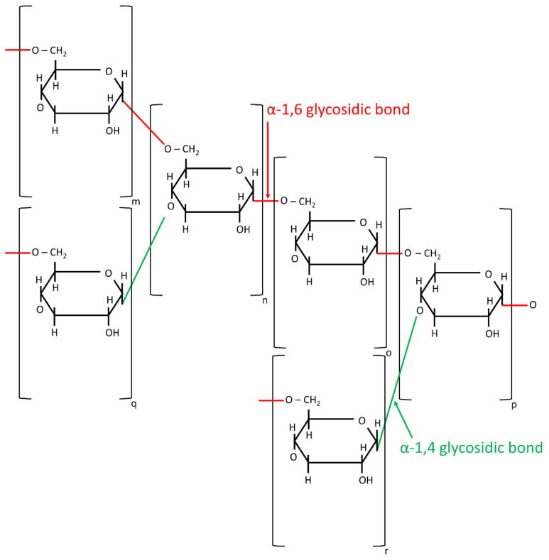
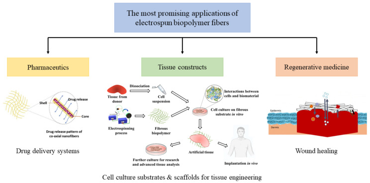
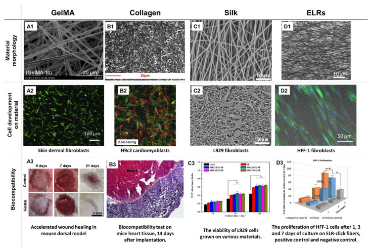
Biopolymers formed into a fibrous morphology through electrospinning are of increasing interest in the field of biomedicine due to their intrinsic biocompatibility and biodegradability and their ability to be biomimetic to various fibrous structures present in animal tissues. However, their mechanical properties are often unsatisfactory and their processing may be troublesome. Thus, extensive research interest is focused on improving these qualities. This review article presents the selection of the recent advances in techniques aimed to improve the electrospinnability of various biopolymers (polysaccharides, polynucleotides, peptides, and phospholipids). The electrospinning of single materials, and the variety of co-polymers, with and without additives, is covered. Additionally, various crosslinking strategies are presented. Examples of cytocompatibility, biocompatibility, and antimicrobial properties are analyzed. Special attention is given to whey protein isolate as an example of a novel, promising, green material with good potential in the field of biomedicine. This review ends with a brief summary and outlook for the biomedical applicability of electrospinnable biopolymers.

Keywords: biopolymers; crosslinking strategies; electrospinning; peptides; tissue engineering.


Conflict of interest statement

There are no conflicts of interest to declare. All authors have approved the final version of the manuscript.

Figures

Figure 1
Figure 1
Two of the most popular electrospinning (ES) setups: needle-based (left) and needleless (right). Setup with rotating mandrils allowing for a high throughput and versatility is depicted in both cases.
Figure 2
Figure 2
The parameters affecting the ES process, segregated by the category.
Figure 3
Figure 3
Impact of various types of chemical crosslinkers on the morphology (A1D1) and chemical stability (A2D2), biocompatibility (A3D3), and 3-day stability (A4D4) of the ES collagen nanofibers. Reprinted from [50] with permission from Elsevier, copyright 2018.
Figure 4
Figure 4
Examples of the optimization studies geared towards identification of the biopolymers’ electrospinnable windows and adequate post-processing. (A) SEM images indicating the relationship between the fraction of the xanthan polymer during processing: (1) 1.5 wt/vol%, (2) 2.0 wt/vol% and (3) 2.5 wt/vol% and the resulting fibrous morphology. Reprinted from [32] with permission from Wiley, copyright 2017. (B) Tertiary diagrams illustrating the correlation between the solvent fraction (acetic and citric acid respectively) in a denatured whole chain collagen (DWCC) polymer system and the morphology of the electrospun products. The diagrams aid in the identification of the optimal solution composition yielding defect-free fiber products. Reprinted from [30] with permission from Elsevier, copyright 2018. (C) The FESEM images presenting the relationship between the concentration of the phospholipids in the initial solution: (1) 35 wt%, (2) 45 wt%, (3) 50 wt% and the fibrous morphology from the resulting polymer product. The significant change in the morphology, from micellar to cylindrical and fibrous, is observed. Reprinted from [40] with permission from Science, copyright 2006. (D) An example of the material electrospun from the salt solution: SEM image of pullulan fibers (15% PUL; 1.0 M NaCl) with crystals of salt visible on the surface. Reprinted from [31] with permission from MDPI, copyright 2017. (E) SEM images of the fibrous morphology stabilized by the introduction of the light-initiated crosslinking—electrospun GelMA fibers prior to UV light (NG-0UV) and after 12 min of UV exposure (NG-12UV). Reprinted from [54] with permission from MDPI, copyright 2019. (F) Diagram illustrating the correlation between the optimal biopolymer concentration and molecular weight and formation of the fibrous products. The optimal guar content with moderate molecular weight ensures fabrication of bead-less fibers; both too high and too low values of MW and concentration yield a mixture of thick fibers and beads. Reprinted from [41] with permission from ACS Omega, copyright 2019. (G) Linear function of the fiber diameter as a function of the biopolymer (xanthan) concentration in the spinning solution. Reprinted from [32] with permission from Wiley, copyright 2017.
Figure 5
Figure 5
A schematic representation of the GelMA fabrication process.
Figure 6
Figure 6
SEM images of WPI-PEO electrospun nanofibers obtained from solutions at: (a) pH 1; (b) pH 7; (c) pH 12, observed under the increasing magnification. Reprinted from [39] with permission from Wiley, copyright 2012.
Figure 7
Figure 7
Scheme of the initial phases of Maillard reaction—the D-glucose molecule. Based on [124].
Figure 8
Figure 8
The scheme of a dextran polymer chain.
Figure 9
Figure 9
The illustration of the most promising applications of electrospun fibrous biopolymer matrices: (Left) Pharmaceutic field as drug delivery system. Drug release pattern of co-axial nanofibers with active compound encapsulated inside the core [10]. (Center) The use of biopolymer fibers as cell culture substrates and scaffolds for tissue engineering (fibrous biopolymer—[10]; culture dish—[134]; interaction between cells and biomaterial—[135]; human posture and Eppendorf—Free sources). (Right) Wound healing application in the field of regenerative medicine. The application of fibrous biopolymer membrane on skin wound and interactions relevant for wound healing that might occur due to porosity of the material, i.e., gas exchange and liquid absorption [8]. Reprinted from [8] with permission from Elsevier, copyright 2020.
Figure 10
Figure 10
The examples of the excellent biocompatibility of the electrospun biopolymeric scaffolds. Electrospun GelMA nanofibers, cross-linked by 10-min UV exposure (A1) and skin dermal fibroblasts on day 7 after seeding on material’s surface (A2). In vivo results revealing an accelerated wound closure efficiency induced by the material (A3). Reprinted from [35] with permission from Elsevier, copyright 2017. Image of electrospun collagen fibers after cross-linking in water with 10% citric acid (B1). Confocal images of H9c2 cardiomyoblasts on the electrospun collagen scaffold after 48 h of cell culture (B2). Image of material biocompatibility towards mice heart tissue 14 days after implantation (B3). Reprinted from [49] with permission from Elsevier, copyright 2015. SEM image of silk nanofibrous matrix with 10% addition (wt/v) of manuka honey, tread with 75% ethanol vapor (v/v) (C1). Image of L929 fibroblast cells on the very same silk nanofibers (also with 10% (wt/v) manuka honey) (C2). The viability of L929 cells grown on different substances used in the research (C3). Reprinted from [111] with permission from Elsevier, copyright 2017. Morphology of electrospun ELR-click fibers (deposition time = 90 s), cross-linked by thermal treatment in water (D1). Phalloidin and DAPI staining of oriented HFF-1 cells on ELR-click fibers (D2). Proliferation histograms of fibroblasts (HFF-1) after 1, 3, and 7 days of culture on ELR-click fibers (FIBERS), positive control, and negative control (D3). Reprinted from [97] with permission from Elsevier, copyright 2018.

References

    1. Schiffman J.D., Schauer C.L. A Review: Electrospinning of Biopolymer Nanofibers and their Applications. Polym. Rev. 2008;48:317–352. doi: 10.1080/15583720802022182. - DOI
    1. Kriegel C., Arrechi A., Kit K., McClements D.J., Weiss J. Fabrication, Functionalization, and Application of Electrospun Biopolymer Nanofibers. Crit. Rev. Food Sci. Nutr. 2008;48:775–797. doi: 10.1080/10408390802241325. - DOI - PubMed
    1. Ng K., Khor H., Hutmacher D. In vitro characterization of natural and synthetic dermal matrices cultured with human dermal fibroblasts. Biomaterials. 2004;25:2807–2818. doi: 10.1016/j.biomaterials.2003.09.058. - DOI - PubMed
    1. Buschle-Diller G., Hawkins A., Cooper J. Electrospun Nanofibers from Biopolymers and Their Biomedical Applications. In: Edwards J.V., Buschle-Diller G., Goheen S.C., editors. Modified Fibers with Medical and Specialty Applications. Springer; Dordrecht, The Netherlands: 2006. pp. 67–80.
    1. Soares R.M., Siqueira N.M., Prabhakaram M.P., Ramakrishna S. Electrospinning and electrospray of bio-based and natural polymers for biomaterials development. Mater. Sci. Eng. C. 2018;92:969–982. doi: 10.1016/j.msec.2018.08.004. - DOI - PubMed