Review
. 2021 Apr 16;13(8):1933.
doi: 10.3390/cancers13081933.

Iatrogenic Ocular Surface Diseases Occurring during and/or after Different Treatments for Ocular Tumours

Affiliations
Review

Iatrogenic Ocular Surface Diseases Occurring during and/or after Different Treatments for Ocular Tumours

Giuseppe Giannaccare et al. Cancers (Basel). .

Abstract

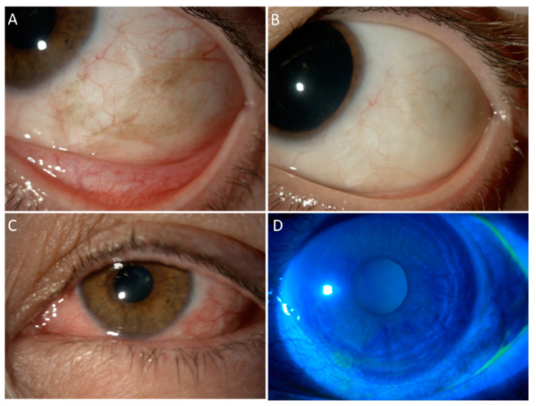
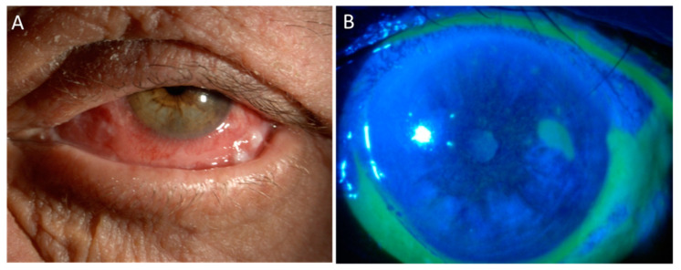
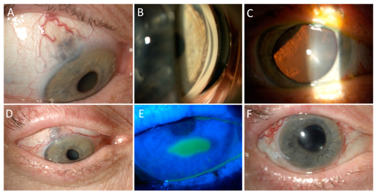
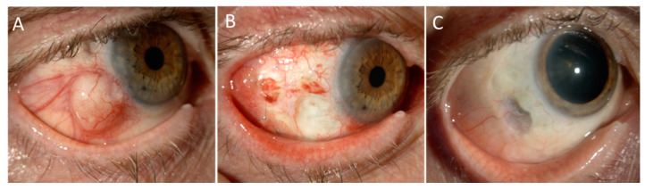
The ocular surface represents a finely regulated system that allows the protection of the eye. It is particularly susceptible to different treatments for intraocular tumours, such as uveal melanoma and conjunctival cancers. Traditionally, the management of ocular tumours depends on the characteristics of the lesion, and is based on a combination of selective surgery, topical chemotherapy, and/or radiotherapy delivered through different mechanisms (e.g., charged-particle radiotherapy or brachytherapy). Possible complications involving the ocular surface range from transient dry eye disease or keratitis up to corneal melting and perforation, which in any case deserve careful evaluation for the risk of permanent sigh-threatening complications. Clinicians involved in the management of these patients must be aware of this risk, in order to reach an early diagnosis and promptly set up an adequate treatment. The present review of the literature will summarize acute and chronic complications affecting the ocular surface following different therapies for the treatment of ocular tumours.

Keywords: anti-cancer agents; choroidal melanoma; complications; dry eye; ocular surface; ocular surface squamous neoplasia; ocular tumours; radiotherapy; uveal melanoma.


Conflict of interest statement

The authors declare no conflict of interest.

Figures

Figure 1
Figure 1
An example of MMC toxicity that successfully resolved with the treatment. A 54-year-old female suffering from biopsy-proven enlarging PAM with moderate atypia in the left eye, which had already been treated with surgical excision and conjunctival graft 8 years earlier for localized PAM with severe atypia. Part (A): Slit lamp picture at baseline, with tumour recurrence surrounding the paralimbal scar of the previous surgery. Part (B): Complete disappearance of pigmented cells 2 years after four courses of MMC 0.02% (one drop QID for 7 days). Part (C): Allergic reaction to MMC starting from the second cycle of treatment, presenting with lid edema, conjunctival swelling, epiphora, and photophobia. Part (D): Fluorescein staining revealed a peripheral superficial corneal epithelial defect. The allergic reaction has been managed with cold compresses, artificial tears, vitamin A ointment, and suspension of MMC. Once the epithelium was completely healed, MMC has been started again in association with weak steroid eye drops and close surveillance.
Figure 2
Figure 2
Toxic blepharoconjunctivitis following MMC. An 80 year-old man affected by relapsing squamous cell carcinoma of the lower fornix in his left eye, who had been treated elsewhere by repeated surgical excisions, presented to our Center after 15 days of continuous treatment with MMC 0.04% QID, with Part (A) severe orbital swelling, erythematous-desquamative blepharitis, and Part (B) corneal epithelial defect and diffuse conjunctival melting with pseudomembranes.
Figure 3
Figure 3
Anterior surface toxicity of PBR. Part (A): A 61 year-old lady presented with a large cilio-choroidal melanoma with scleral and iris invasion in the right eye, which was her only eye, as the left one had been previously enucleated due to a trauma. Part (B): Circumferential invasion of the iris angle by pigmented cells was detected on gonioscopy. Part (C): Sectorial cataract was present due to lens infiltration by the melanoma, as well as inferior exudative retinal detachment. The patient was treated with PBR sectorial irradiation of the ciliary body, anterior choroid, and whole iris. Harvesting of limbal stem cell was not performed due to the extraocular extension. Part (D): Three months after treatment, the patient developed madarosis and scarring of the superior eyelid and diffuse punctate keratitis that was managed with the regular use of artificial tears and vitamin A ointment in association with atropine and unpreserved mild steroids. Part (E): Eight months after treatment, a neurotrophic keratopathy developed and was treated with gas-permeable contact lenses and hourly tear substitutes. Part (F): Two years after PBR, the tumour has regressed to a flat scar. The patient has undergone cataract surgery and anti-vascular endothelial growth factor injections, vitrectomy, and endolaser for neovascular glaucoma due to ischemic retinopathy, with a residual visual acuity of 20/200 due to radiation maculopathy.
Figure 4
Figure 4
Ruthenium brachytheraphy for invasive squamous cell carcinoma of the conjunctiva. Part (A): A 71 year-old man affected by invasive squamous cell carcinoma of the conjunctiva in the right eye that aggressively recurred shortly after a partial excision with scleral lamellectomy and cryotherapy done at a local hospital. In the attempt to save the globe, he was treated with surgical excision, Ruthenium-106 brachitherapy, and ocular surface reconstruction with amniotic membrane graft. Part (B): One-month after surgery, the amniotic membrane had been completely reabsorbed and the patient was treated with two courses of adjuvant 5-FU and intensive topical lubrification and soft steroids with slow tapering. Part (C): Two years after treatment, the eye is preserved with a 20/20 vision with no signs of recurrence. Note the scleral thinning, well covered by tenon and conjunctiva, in the area of previous full-thickness tumour invasion. This area is being monitored by means of anterior segment optical coherence tomography to exclude its evolution. In such case, a wide scleral patch would be indicated.

Similar articles

Cited by

References

    1. Singh A.D., Seregard S., editors. Ocular Tumours. Volume 7. S. Karger AG; Basel, Switzerland: 2016. (ESASO Course Series).
    1. Singh A.D., Turell M.E., Topham A.K. Uveal melanoma: Trends in incidence, treatment, and survival. Ophthalmology. 2011;118:1881–1885. doi: 10.1016/j.ophtha.2011.01.040. - DOI - PubMed
    1. Virgili G., Gatta G., Ciccolallo L., Capocaccia R., Biggeri A., Crocetti E., Lutz J.M., Paci E. Incidence of Uveal Melanoma in Europe. Ophthalmology. 2007;114:2309–2315.e2. doi: 10.1016/j.ophtha.2007.01.032. - DOI - PubMed
    1. Hammer H., Oláh J., Tóth-Molnár E. Dysplastic nevi are a risk factor for uveal melanoma. Eur. J. Ophthalmol. 1996;6:472–474. doi: 10.1177/112067219600600423. - DOI - PubMed
    1. Eskelin S., Kivelä T. Mode of presentation and time to treatment of uveal melanoma in Finland. Br. J. Ophthalmol. 2002;86:333–338. doi: 10.1136/bjo.86.3.333. - DOI - PMC - PubMed

LinkOut - more resources