Review
. 2021 Apr 20;13(4):709.
doi: 10.3390/v13040709.

Insights into the Pathogenesis of Viral Haemorrhagic Fever Based on Virus Tropism and Tissue Lesions of Natural Rift Valley Fever

Affiliations
Review

Insights into the Pathogenesis of Viral Haemorrhagic Fever Based on Virus Tropism and Tissue Lesions of Natural Rift Valley Fever

Lieza Odendaal et al. Viruses. .

Abstract

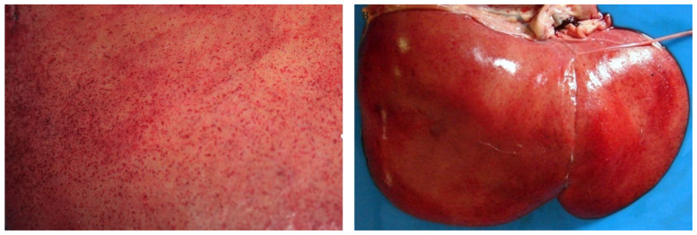
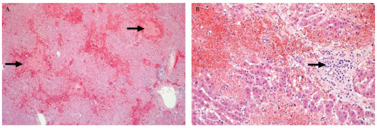
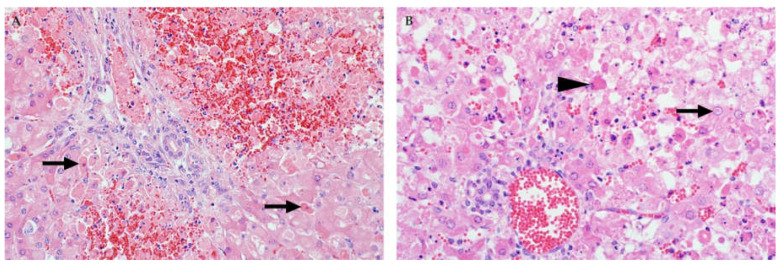
Rift Valley fever phlebovirus (RVFV) infects humans and a wide range of ungulates and historically has caused devastating epidemics in Africa and the Arabian Peninsula. Lesions of naturally infected cases of Rift Valley fever (RVF) have only been described in detail in sheep with a few reports concerning cattle and humans. The most frequently observed lesion in both ruminants and humans is randomly distributed necrosis, particularly in the liver. Lesions supportive of vascular endothelial injury are also present and include mild hydropericardium, hydrothorax and ascites; marked pulmonary congestion and oedema; lymph node congestion and oedema; and haemorrhages in many tissues. Although a complete understanding of RVF pathogenesis is still lacking, antigen-presenting cells in the skin are likely the early targets of the virus. Following suppression of type I IFN production and necrosis of dermal cells, RVFV spreads systemically, resulting in infection and necrosis of other cells in a variety of organs. Failure of both the innate and adaptive immune responses to control infection is exacerbated by apoptosis of lymphocytes. An excessive pro-inflammatory cytokine and chemokine response leads to microcirculatory dysfunction. Additionally, impairment of the coagulation system results in widespread haemorrhages. Fatal outcomes result from multiorgan failure, oedema in many organs (including the lungs and brain), hypotension, and circulatory shock. Here, we summarize current understanding of RVF cellular tropism as informed by lesions caused by natural infections. We specifically examine how extant knowledge informs current understanding regarding pathogenesis of the haemorrhagic fever form of RVF, identifying opportunities for future research.

Keywords: Bunyavirales; Rift Valley fever phlebovirus; arbovirus; emerging diseases; pathogenesis; pathology; tropism; zoonotic disease.


Conflict of interest statement

The authors declare no conflict of interest.

Figures

Figure 1
Figure 1
Macroscopic pathology of the liver in sheep naturally infected with RVFV. (A) Innumerable petechia on the parietal surface of the liver of an adult sheep. (B) Liver of a new-born lamb with extensive necrosis and pinpoint subcapsular petechia giving the liver a pale yellow to red mottled appearance.
Figure 2
Figure 2
Histopathology of the liver of cattle naturally infected with RVFV (Haematoxylin and eosin (H&E) stain). (A) Random multifocal to coalescing necrosis and haemorrhage (arrows), original magnification (mag) 40×. (B) Necrosis of hepatocytes extends into the periportal zones, specifically affecting hepatocytes of the limiting plate. Mild mononuclear cell inflammation (arrow) is also present in the portal area, mag 200×.
Figure 3
Figure 3
Histopathology of the liver in young lambs naturally infected with RVFV (H&E stain). (A) Hepatocytes with features of apoptosis (arrows), characterized by disassociation of cells, hypereosinophilic cytoplasm and pyknosis or karyorrhexis, mag 400×. (B) Diffuse necrosis of hepatocytes, filamentous eosinophilic intranuclear inclusions (arrow), and late apoptotic bodies (arrowhead), magnification 600×.
Figure 4
Figure 4
Histopathology of the liver in sheep foetuses naturally infected with RVFV. (A) Multiple foci of liquefactive hepatic necrosis (arrows), also referred to as primary foci, are present (H&E stain). There is also severe periportal oedema (arrowhead), mag 40×. (B) Nuclear fragments and remnants of reticulin fibres (arrow) are present in an area of liquefactive necrosis (Gordon and Sweets’ silver stain for reticular fibres with Van Gieson counterstain), mag 400×.
Figure 5
Figure 5
Immunolabelling for RVFV in the liver of sheep (polyclonal mouse anti-RVFV nucleoprotein antibody, avidin-biotin-peroxidase detection system, NovaRED chromogen and haematoxylin counterstain). (A) In this specimen from an adult sheep, viral antigen (arrow) is present in multiple foci in the lobule. Labelling is present in the cytoplasm of injured hepatocytes and is diffuse or fine granular, mag 400×. (B) In young lambs, viral antigen is diffusely present in the cytoplasm of hepatocytes, but labelling is sparse in primary foci (arrows), mag 100×.
Figure 6
Figure 6
Kidney of RVFV-infected adult sheep (Jones’ methenamine silver stain). (A) Lesions in necrotic tubules are characterized by tubular epithelial cell pyknosis, karyorrhexis, and karyolysis accompanied by detachment of the epithelium from the basement membrane (arrow), mag 200×. (B) Pyknosis and karyorrhexis are present in a renal glomerulus (arrow) with a marked decrease in mesangial cellularity. Pyknosis is also present in the interstitium (arrowhead) and in many tubular epithelial cells, mag 400×.
Figure 7
Figure 7
Immunolabelling for RVFV in the kidney of sheep (NovaRed IHC as detailed in Figure 5). (A) In this specimen from a young lamb, viral antigen is prominent in Lacis cells in the glomerulus (arrow). Other cells, morphologically consistent with endothelial cells also labelled in the glomerulus. Cells in the macula densa are not labelled, mag 400×. (B) Adult sheep with immunolabelling of necrotic renal tubular epithelial cells (arrow) in the renal cortex, mag 600×.
Figure 8
Figure 8
Spleen of an RVFV-infected adult sheep. (A) Scattered sub-capsular petechiae are present. (B) Lymphocytolysis is most apparent in the germinal centre (arrow) (H&E stain), mag 200×.
Figure 9
Figure 9
Spleen of an RVFV-infected adult sheep with sequential sections immunolabelled for T and B lymphocytes respectively (polyclonal rabbit anti-CD3 and anti-CD20 antibodies, micro-polymer detection system, DAB chromogen and haematoxylin counterstain). (A) Labelling with the anti-CD3 antibody shows a marked loss of T lymphocytes. Only scattered T lymphocytes remain in the red pulp and there is severe depletion of the periarteriolar lymph sheath (arrow), mag 100×. Inset: Spleen from a healthy control sheep showing a normal distribution of abundant T lymphocytes, mag 100×. (B) Labelling with the anti-CD 20 antibody shows necrotic debris in the germinal centre (arrow) with a few residual B lymphocytes in the marginal zones (arrowhead) of the periarteriolar lymph sheath and in the red pulp, mag 100×. Inset: Spleen from a healthy sheep with multiple lymphoid follicles that contain many B lymphocytes, mag 100×.
Figure 10
Figure 10
Spleen of a RVFV-infected sheep (NovaRed IHC as detailed in Figure 5). (A) In this specimen from an adult sheep, viral antigen is present in multiple macrophages in the marginal zone of the white pulp (arrow). Labelling is also present in a tingible-body macrophage in the white pulp (arrowhead), mag 400×. (B) Prominent immunolabelling of non-cell-associated antigen and cellular debris in the subcapsular red pulp (arrow). Viral antigen is also present in endothelial cells (arrowhead) and the capsular smooth myocytes, mag 200×.
Figure 11
Figure 11
Lungs of RVFV-infected lamb and adult sheep. (A) The interstitium in the lungs of this lamb is markedly expanded due to oedema, and dark red areas of atelectasis are present (arrow). Multifocal petechiae are also present in the serosa. (B) Microscopically, intra-alveolar (arrow) and interstitial (arrowhead) lung oedema is present in this adult sheep (H&E stain), mag 100×.
Figure 12
Figure 12
Heart of a RVFV-infected sheep (NovaRed IHC as detailed in Figure 5). (A) In this specimen from a young lamb, viral antigen is present in endothelial cells (arrow) and cardiomyocytes (arrowhead), mag 400×. (B) Immunolabelling is especially prominent in the Purkinje fibres (arrow), mag 100×. Inset: Negative control using a following slide from the same case (avidin-biotin-peroxidase system using mouse polyclonal anti-Wesselsbron antibody, NovaRED peroxidase substrate with haematoxylin counterstain), mag 100×.
Figure 13
Figure 13
Omentum and rumen of an RVFV-infected adult sheep. (A) Marked congestion of the omental blood vessels with petechiae along the vessels and in the omental fat (arrow). (B) Petechiae and ecchymoses in the serosa of the rumen (arrow).
Figure 14
Figure 14
Skin and brain of RVFV-infected sheep (NovaRed IHC as detailed in Figure 5). (A) Immunolabelling in the epidermis (arrow) of an adult sheep, mag ×400. (B) Viral antigen in vascular endothelial cells and cellular debris in small blood vessels in the meninges (arrow) of a young lamb, mag 200×.
Figure 15
Figure 15
Placenta of RVFV-infected sheep. (A) Diffuse necrosis of trophoblasts with cellular debris (arrow) between the villi (H&E stain), mag 100×. (B) Immunolabelling in trophoblasts and cellular debris (arrow) in the cotyledonary chorioallantois (NovaRed IHC as detailed in Figure 5), mag 400×.
Figure 16
Figure 16
Model of the pathogenic mechanism underlying RVFV infection. Following a mosquito bite, the virus is endocytosed by antigen-presenting cells. Suppression of type I IFN production and necrosis of infected macrophages and dendritic cells cause wide dissemination of viruses. This systemic spread leads to necrosis in a variety of tissues and cells together with suppression of both the innate and adaptive immune responses. Apoptosis of lymphocytes might occur through mediator effects and loss of dendritic cell support, exacerbating the failure of the immune response. An excessive pro-inflammatory cytokine and chemokine response follow, resulting in increased microcirculatory dysfunction through the action of inflammatory mediators. Impairment of the coagulation system results in widespread haemorrhages. Fatal outcomes result from multiorgan failure, oedema in many organs (including the lungs and brain), hypotension and circulatory shock. DC, dendritic cell. NO, nitric oxide. (Illustration adapted from Bray M, 2005).

Similar articles

Cited by

References

    1. Pepin M., Bouloy M., Bird B.H., Kemp A., Paweska J. Rift Valley fever virus (Bunyaviridae: Phlebovirus): An update on pathogenesis, molecular epidemiology, vectors, diagnostics and prevention. Vet. Res. 2010;41:e2010033. doi: 10.1051/vetres/2010033. - DOI - PMC - PubMed
    1. Abudurexiti A., Adkins S., Alioto D., Alkhovsky S.V., Avsic-Zupanc T., Ballinger M.J., Bente D.A., Beer M., Bergeron E., Blair C.D., et al. Taxonomy of the order Bunyavirales: Update 2019. Arch. Virol. 2019;164:1949–1965. doi: 10.1007/s00705-019-04253-6. - DOI - PMC - PubMed
    1. Davies F.G., Linthicum K.J., James A.D. Rainfall and epizootic Rift Valley fever. Bull. World Health Organ. 1985;63:941–943. - PMC - PubMed
    1. Anyangu A.S., Gould L.H., Sharif S.K., Nguku P.M., Omolo J.O., Mutonga D., Rao C.Y., Lederman E.R., Schnabel D., Paweska J.T., et al. Risk factors for severe Rift Valley fever infection in Kenya, 2007. Am. J. Trop. Med. Hyg. 2010;83(Suppl. 2):14–21. doi: 10.4269/ajtmh.2010.09-0293. - DOI - PMC - PubMed
    1. Madani T.A., Al-Mazrou Y.Y., Al-Jeffri M.H., Mishkhas A.A., Al-Rabeah A.M., Turkistani A.M., Al-Sayed M.O., Abodahish A.A., Khan A.S., Ksiazek T.G., et al. Rift Valley fever epidemic in Saudi Arabia: Epidemiological, clinical, and laboratory characteristics. Clin. Infect. Dis. 2003;37:1084–1092. doi: 10.1086/378747. - DOI - PubMed

LinkOut - more resources