Review
. 2021 Apr 20;22(8):4252.
doi: 10.3390/ijms22084252.

Small Hsps as Therapeutic Targets of Cystic Fibrosis Transmembrane Conductance Regulator Protein

Affiliations
Review

Small Hsps as Therapeutic Targets of Cystic Fibrosis Transmembrane Conductance Regulator Protein

Stéphanie Simon et al. Int J Mol Sci. .

Abstract

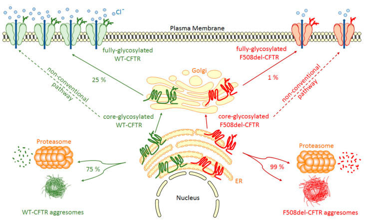
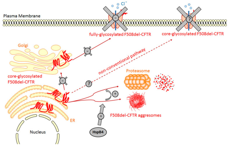
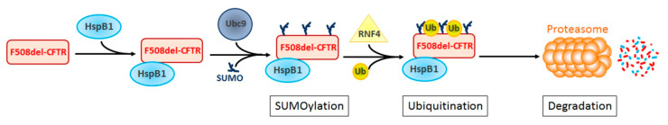
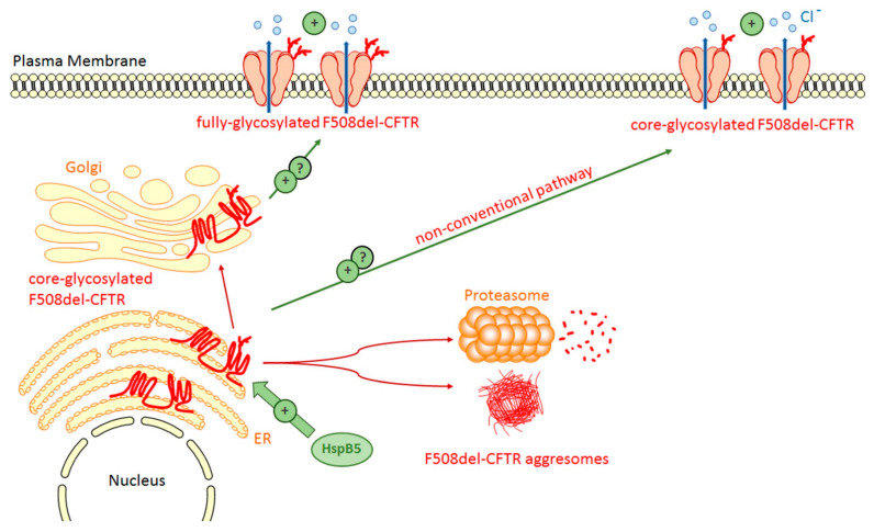
Human small heat shock proteins are molecular chaperones that regulate fundamental cellular processes in normal and pathological cells. Here, we have reviewed the role played by HspB1, HspB4 and HspB5 in the context of Cystic Fibrosis (CF), a severe monogenic autosomal recessive disease linked to mutations in Cystic Fibrosis Transmembrane conductance Regulator protein (CFTR) some of which trigger its misfolding and rapid degradation, particularly the most frequent one, F508del-CFTR. While HspB1 and HspB4 favor the degradation of CFTR mutants, HspB5 and particularly one of its phosphorylated forms positively enhance the transport at the plasma membrane, stability and function of the CFTR mutant. Moreover, HspB5 molecules stimulate the cellular efficiency of currently used CF therapeutic molecules. Different strategies are suggested to modulate the level of expression or the activity of these small heat shock proteins in view of potential in vivo therapeutic approaches. We then conclude with other small heat shock proteins that should be tested or further studied to improve our knowledge of CFTR processing.

Keywords: CFTR; HspB1; HspB4; HspB5; cystic fibrosis; sHsps.


Conflict of interest statement

The authors declare no conflict of interest.

Figures

Figure 1
Figure 1
WT- and F508del-CFTR biogenesis. After synthesis in the Endoplasmic Reticulum (ER), the main part of core-glycosylated CFTR can form aggresomes or be recognized by the Endoplasmic Reticulum Quality Control (ERQC) system and directed to the proteasome to be degraded. CFTR escaped from ERQC undergoes different steps of glycosylation in the Golgi before being transported to the plasma membrane where it regulates ion channels. F508del-CFTR at the plasma membrane is less abundant and has only a low activity compared to WT-CFTR. A non-conventional trafficking of the CFTR protein can be activated when the conventional trafficking is blocked allowing expression of core-glycosylated CFTR at the plasma membrane.
Figure 2
Figure 2
HspB4 promotes the degradation of F508del-CFTR. The transient expression of HspB4 reveals that it is able to bind F508del-CFTR. In vitro experiments have revealed that, through the binding of the NDB1 domain, HspB4 probably maintains F508del-CFTR in a soluble form that facilitates its degradation by the proteasome.
Figure 3
Figure 3
HspB1 promotes the degradation by the proteasome of F508del-CFTR through SUMO and ubiquitin pathways. HspB1 interacts specifically with the NDB1 domain of F508del-CFTR. This interaction favors the recruitment of UBC9 and the subsequent SUMOylation of F508del-CFTR. SUMOylated F508del-CFTR-HspB1 complex is recognized by the ubiquitin ligase RNF4, known to ubiquitinate SUMOylated proteins, thus causing their degradation by the proteasome.
Figure 4
Figure 4
HspB5 favors the rescue of F508del-CFTR. The transient expression of HspB5 increases the stability of core-glycosylated F508del-CFTR thus enhancing the level of F508del-CFTR at the plasma membrane and its activity. The type of glycosylated F508del-CFTR form at the plasma membrane is currently not determined. One phosphomimic mutant of HspB5 (S19D, S45A, S59D) is more effective than wild-type HspB5 to correct F508del-CFTR.

Similar articles

Cited by

References

    1. Riordan J.R., Rommens J.M., Kerem B., Alon N., Rozmahel R., Grzelczak Z., Zielenski J., Lok S., Plavsic N., Chou J.L. Identification of the Cystic Fibrosis Gene: Cloning and Characterization of Complementary DNA. Science. 1989;245:1066–1073. doi: 10.1126/science.2475911. - DOI - PubMed
    1. Hyde S.C., Emsley P., Hartshorn M.J., Mimmack M.M., Gileadi U., Pearce S.R., Gallagher M.P., Gill D.R., Hubbard R.E., Higgins C.F. Structural Model of ATP-Binding Proteins Associated with Cystic Fibrosis, Multidrug Resistance and Bacterial Transport. Nature. 1990;346:362–365. doi: 10.1038/346362a0. - DOI - PubMed
    1. Zhang Z., Liu F., Chen J. Conformational Changes of CFTR upon Phosphorylation and ATP Binding. Cell. 2017;170:483–491.e8. doi: 10.1016/j.cell.2017.06.041. - DOI - PubMed
    1. Hwang T.-C., Sheppard D.N. Gating of the CFTR Cl- Channel by ATP-Driven Nucleotide-Binding Domain Dimerisation. J. Physiol. 2009;587:2151–2161. doi: 10.1113/jphysiol.2009.171595. - DOI - PMC - PubMed
    1. Crawford I., Maloney P.C., Zeitlin P.L., Guggino W.B., Hyde S.C., Turley H., Gatter K.C., Harris A., Higgins C.F. Immunocytochemical Localization of the Cystic Fibrosis Gene Product CFTR. Proc. Natl. Acad. Sci. USA. 1991;88:9262–9266. doi: 10.1073/pnas.88.20.9262. - DOI - PMC - PubMed

MeSH terms