

Review
doi: 10.3390/ijms22084270.
Revisiting the Impact of Local Leptin Signaling in Folliculogenesis and Oocyte Maturation in Obese Mothers
Affiliations
- PMID: 33924072
- PMCID: PMC8074257
- DOI: 10.3390/ijms22084270
Item in Clipboard
Review
Revisiting the Impact of Local Leptin Signaling in Folliculogenesis and Oocyte Maturation in Obese Mothers
Int J Mol Sci.
.
Abstract
The complex nature of folliculogenesis regulation accounts for its susceptibility to maternal physiological fitness. In obese mothers, progressive expansion of adipose tissue culminates with severe hyperestrogenism and hyperleptinemia with detrimental effects for ovarian performance. Indeed, maternal obesity is associated with the establishment of ovarian leptin resistance. This review summarizes current knowledge on potential effects of impaired leptin signaling throughout folliculogenesis and oocyte developmental competence in mice and women.
Keywords: folliculogenesis; leptin; obesity; oocyte; ovary.
Conflict of interest statement
The authors declare no conflict of interest.
Figures
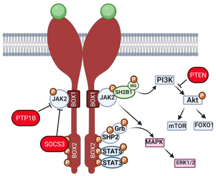
Schematic representation of leptin signaling pathway cascade. Leptin binds to its dimerized membrane receptor, and signal propagation starts. Janus kinase 2 (JAK2) phosphorylation causes transition of phosphate groups to three tyrosines within BOX2 of ObR and activation of (i) SH2-domain containing protein tyrosine phosphatase (SHP-2), which, in turns, binds its adapter molecule Grb-2 and activates downstream signaling, resulting in extracellular signal regulated kinase (ERK) 1/2 activation, (ii) signal transducer and activator of transcription (STAT) 5 activation and (iii) STAT3 activation. The phosphorylation of JAK2 also activates the MAPK signaling pathway and promotes SH2B adaptor protein 1 (SH2B1) and insulin receptor substrate (IRS) binding, which initiates the phosphatidylinositol 3 kinase (PI3K) pathway, which leads to phosphorylation of protein kinase B (Akt), mammalian target of rapamycin (mTOR) and forkhead box O1 (FOXO1) activation. During hyperactivation of the leptin signaling pathway, two main inhibitors can be transcribed—protein tyrosine phosphatase (PTP) 1B, which dephosphorylates JAK2 and suppressor of cytokine signaling (SOCS) 3, which blocks tyrosine and JAK2 phosphorylation. Created with BioRender.com , accessed on 1 November 2020.
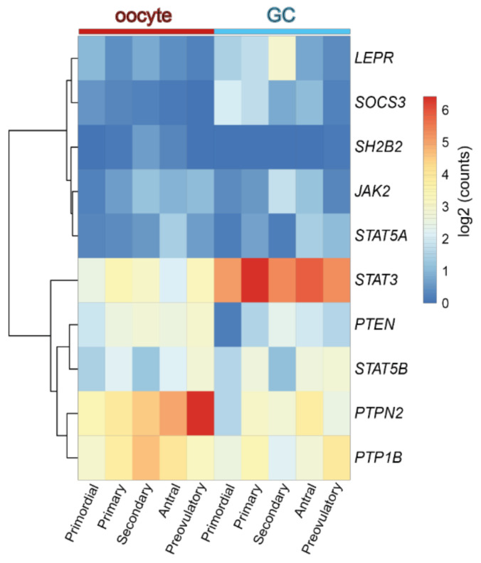
Heatmap representing the expression level of transcripts from the leptin signaling pathway components in human oocyte and granulosa cells (GC) throughout folliculogenesis. Primordial = primordial follicle; Primary = primary follicle; Secondary = secondary follicle; Antral = antral follicle; Preovulatory = preovulatory follicle. Color code from blue to red indicates the relative gene expression level from low to high, respectively. Data from Zhang et al. 2018 [30]. Leptin receptor (LEPR), suppressor of cytokine signaling 3 (SOCS3), SH2B Adaptor Protein 1 (SH2B1), Janus kinase 2 (JAK2), signal transducer and activator of transcription (STAT), protein tyrosine phosphatase non-receptor type 2 (PTPN2), protein tyrosine phosphatase (PTP) 1B.
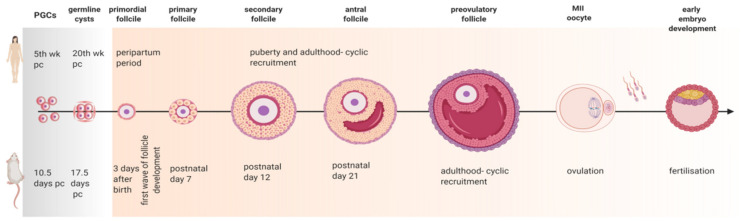
Diagram of folliculogenesis in mice and women. Approximately five weeks (wk) post coitum (pc) in women and 10.5 days (d) pc in mice, primordial germ cells (PGC) arrive at the genital ridge. Germline cysts start breaking 15 wk later in women and 7 days later in mice, creating the primordial follicles. During the peripartum period in woman, and within 3 days after birth in mice, primordial follicles are created. The process of follicle development in women is asynchronous, with menstrual cyclicity being started at puberty. In mice, the first wave of follicle development is detected around postnatal day 7, when the first primary follicles are originated, followed by secondary follicles detected at postnatal day 12 and early antral follicles around postnatal day 21. When mice reach sexual maturity, cyclic recruitment of follicles begins. After ovulation metaphase II (MII) oocyte is released into the oviduct where fertilization takes place, followed by early embryo development. Created with BioRender.com , accessed on 1 November 2020.
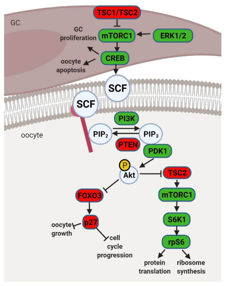
Schematic representation of mammalian target of rapamycin (mTOR) and phosphatidylinositol 3 kinase (PI3K) signaling pathway and its downstream regulators in the granulosa cells (GC) and oocyte. Extracellular signal-regulated protein kinase ½ (ERK1/2) activates mTOR complex 1 (mTORC1) in pre-GC to initiate the activation of primordial follicles. mTORC1 activates cyclic AMP-response element binding protein (CREB), which promotes stem cell factor (SCF) transcription and stimulates PI3K signaling but also affects pre-GC proliferation and oocyte apoptosis. mTOR signaling negative regulators TSC1 and TSC2 suppress mTORC1 activity. PI3K signaling pathway is activated by SCF in the oocyte. PI3K phosphorylates phosphatidylinositol-4,5-biphosphate (PIP2) to phosphatidylinositol-3,4,5-triphosphate (PIP3), which interacts with 3-phosphoinositide dependent protein kinase 1 (PDK1) for subsequent phosphorylation of protein kinase B (Akt) and forkhead box O3 (FOXO3) with its downstream mediator, cyclin-dependent kinase inhibitor 1B (p27). Upon phosphorylation, the inhibitory effect of FOXO3 and p27 on cell cycle progression and oocyte growth is inhibited, and the primordial follicle is recruited. Akt also activates ribosomal protein S6 kinase beta-1 (S6K1) through inhibition of TSC2 with subsequent mTORC1 activation and further ribosomal protein S6 (rpS6) phosphorylation, which leads to protein translation and ribosome synthesis. Factors indicated in red are associated with follicle dormancy; molecules indicated in green are associated with follicle activation. Created with BioRender.com , accessed on 1 November 2020.
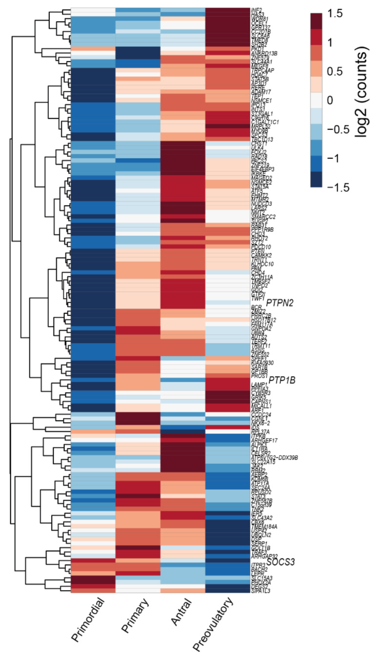
Heatmap representing transcription profile of genes identified as differentially expressed in cumulus cells of mice both fed high fat diet for 4 wk and treated with leptin (for details please see [6]) in human granulosa cells throughout folliculogenesis. Primordial = primordial follicle; Primary = primary follicle; Antral = antral follicle; Preovulatory = preovulatory follicle. Color code from blue to red indicates the relative gene expression level from low to high, respectively. Data from Zhang et al. 2018 [30]; 134 DEGs plus 10 leptin pathway genes—Table S2. Suppressor of cytokine signaling (SOCS) 3, protein tyrosine phosphatase non-receptor type 2 (PTPN2), protein tyrosine phosphatase (PTP) 1B.
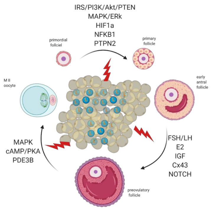
Adipose tissue secretes excessive amounts of leptin (L) during obesity. Leptin signaling in the ovaries of obese mothers is altered, culminating with the establishment of leptin resistance. As a result, signaling pathways governing primordial follicle activation, preovulatory follicle formation, and oocyte maturation can be affected. Created with BioRender.com , accessed on 1 November 2020.
References
-
- Yang X., Wu L.L., Chura L.R., Liang X., Lane M., Norman R.J., Robker R.L. Exposure to lipid-rich follicular fluid is associated with endoplasmic reticulum stress and impaired oocyte maturation in cumulus-oocyte complexes. Fertil. Steril. 2012;97:1438–1443. doi: 10.1016/j.fertnstert.2012.02.034. - DOI - PubMed
Publication types
MeSH terms
Substances
Grants and funding
LinkOut - more resources
Full Text Sources
Other Literature Sources
Medical
