. 2021 Apr 13;22(8):3996.
doi: 10.3390/ijms22083996.

In Vitro Efficacy of Bacterial Cellulose Dressings Chemisorbed with Antiseptics against Biofilm Formed by Pathogens Isolated from Chronic Wounds

Affiliations

In Vitro Efficacy of Bacterial Cellulose Dressings Chemisorbed with Antiseptics against Biofilm Formed by Pathogens Isolated from Chronic Wounds

Karolina Dydak et al. Int J Mol Sci. .

Abstract

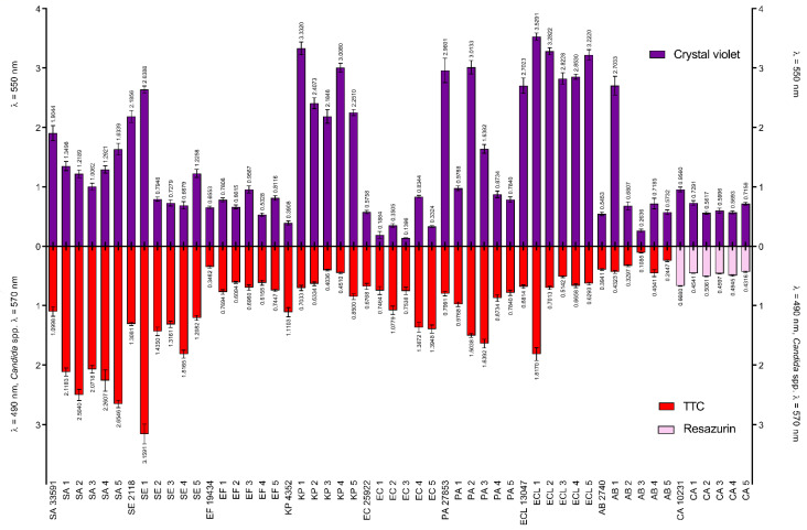
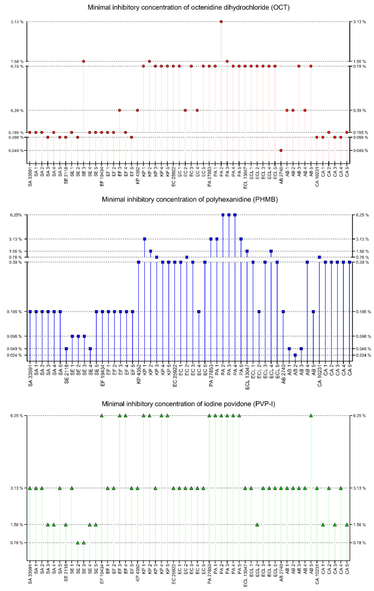
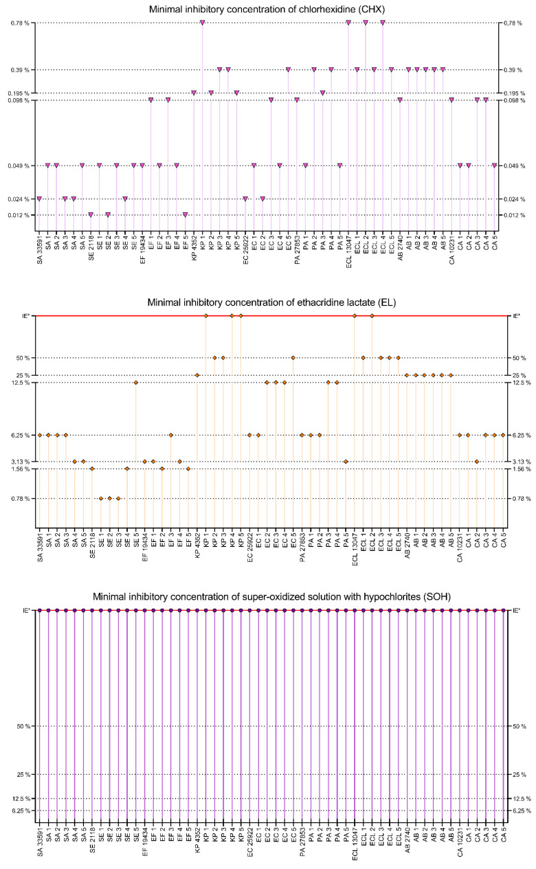
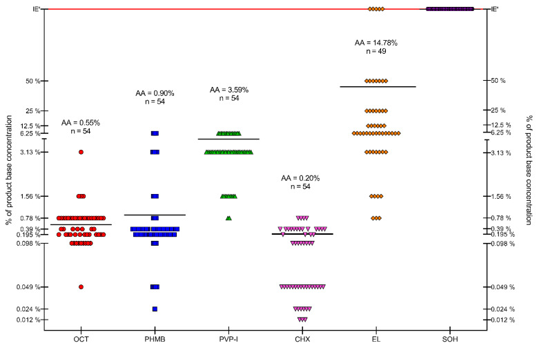
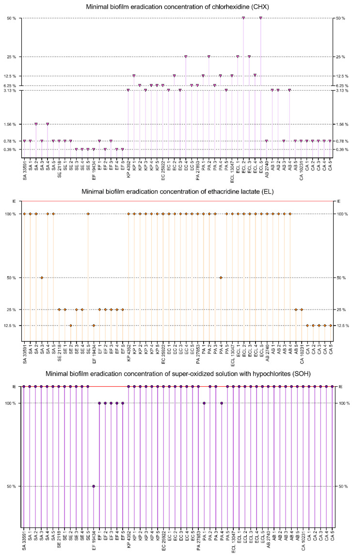
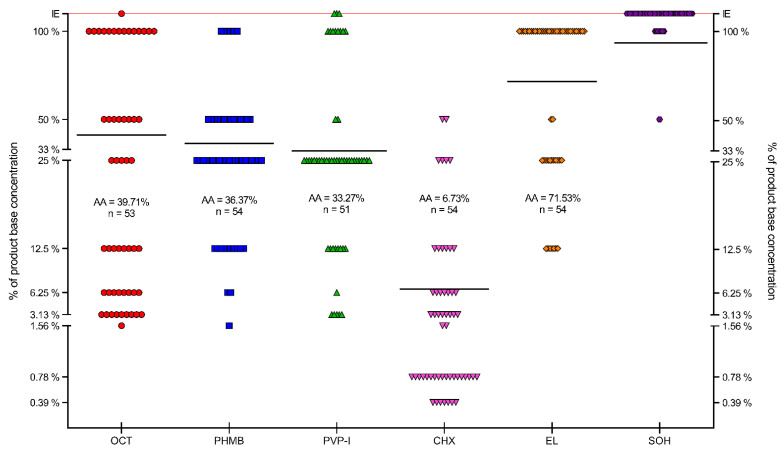
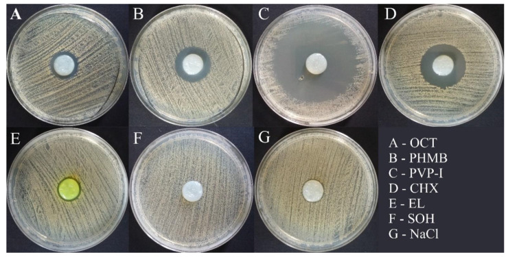
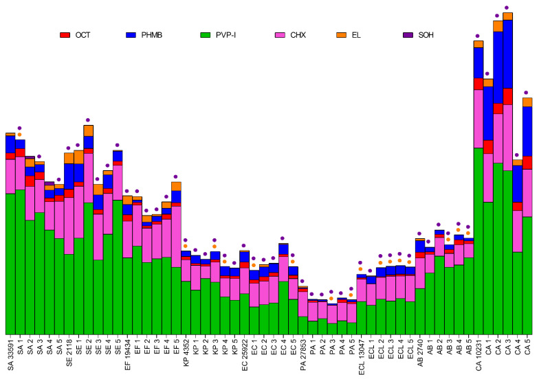
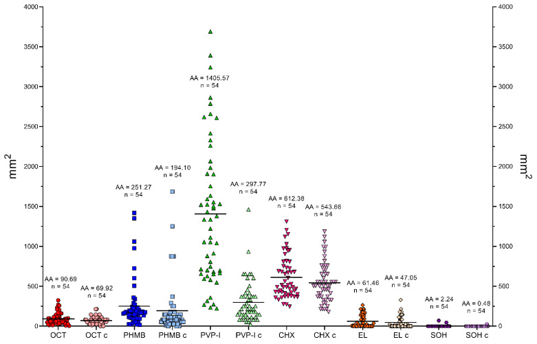
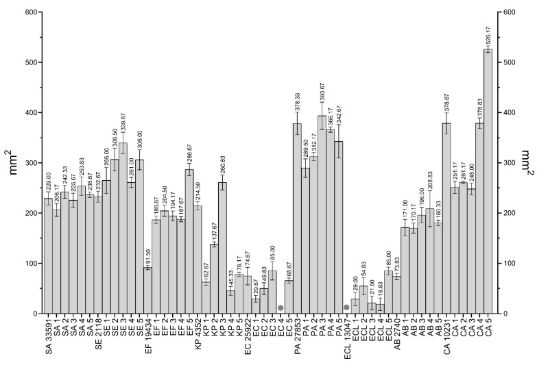
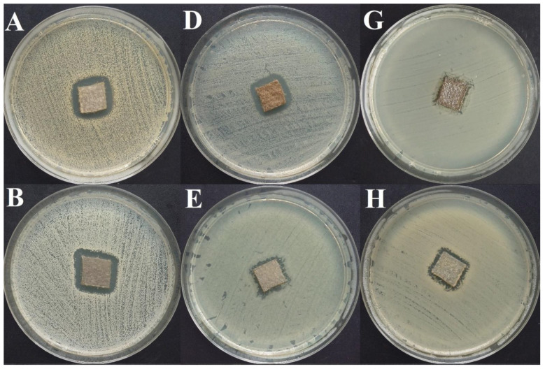
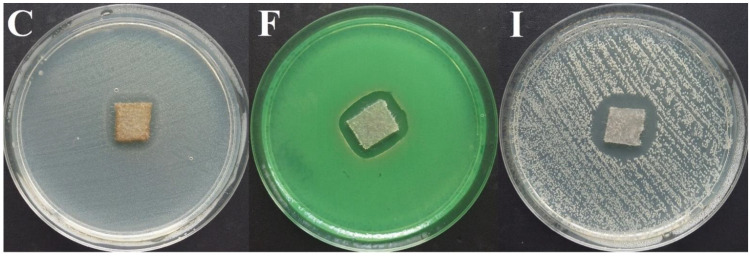
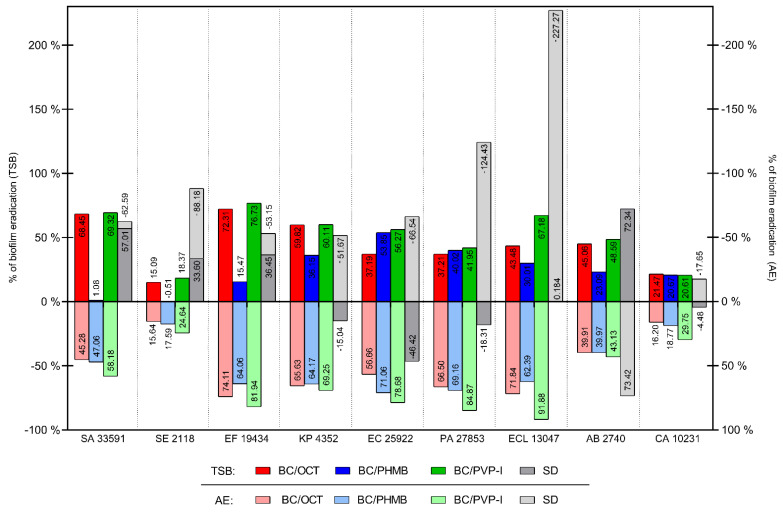
Local administration of antiseptics is required to prevent and fight against biofilm-based infections of chronic wounds. One of the methods used for delivering antiseptics to infected wounds is the application of dressings chemisorbed with antimicrobials. Dressings made of bacterial cellulose (BC) display several features, making them suitable for such a purpose. This work aimed to compare the activity of commonly used antiseptic molecules: octenidine, polyhexanide, povidone-iodine, chlorhexidine, ethacridine lactate, and hypochlorous solutions and to evaluate their usefulness as active substances of BC dressings against 48 bacterial strains (8 species) and 6 yeast strains (1 species). A silver dressing was applied as a control material of proven antimicrobial activity. The methodology applied included the assessment of minimal inhibitory concentrations (MIC) and minimal biofilm eradication concentration (MBEC), the modified disc-diffusion method, and the modified antibiofilm dressing activity measurement (A.D.A.M.) method. While in 96-well plate-based methods (MIC and MBEC assessment), the highest antimicrobial activity was recorded for chlorhexidine, in the modified disc-diffusion method and in the modified A.D.A.M test, povidone-iodine performed the best. In an in vitro setting simulating chronic wound conditions, BC dressings chemisorbed with polyhexanide, octenidine, or povidone-iodine displayed a similar or even higher antibiofilm activity than the control dressing containing silver molecules. If translated into clinical conditions, the obtained results suggest high applicability of BC dressings chemisorbed with antiseptics to eradicate biofilm from chronic wounds.

Keywords: antiseptics; bacterial cellulose; chronic wounds; dressing.


Conflict of interest statement

The authors declare no conflict of interest.

Figures

Figure 1
Figure 1
Stages of wound infection and corresponding clinical symptoms [9,10,12]. * antisepsis, wound debridement, antimicrobial dressings, non-antibiotic local treatment. Local antibiotic therapy is not recommended. ** systemic empiric antibiotic therapy depends on the clinical condition of the patient. Empirical treatment should consider the expected pathogens, the site and nature of the infection, and cover the broadest possible spectrum of microorganisms. Empirical antibiotic therapy should be turned into targeted antibiotic therapy as soon as possible after obtaining microbiology test results.
Figure 2
Figure 2
Examples of commercially available modern active dressings and their applications [84,86,87,88,89,90,91,92,93,94,95,96]. PHMB—polyhexanide; PVP-I—povidone-iodine; CHX—chlorhexidine; OCT—octenidine.
Figure 3
Figure 3
Examples of compounds/substances/molecules introduced to bacterial cellulose (BC) as an antimicrobial additive. MgO—magnesium oxide; CuO—copper oxide; TiO2—titanium dioxide; ZnO—zinc oxide; Na-MMT—sodium montmorillonite; Ca-MMT—calcium montmorillonite; Cu-MMT—copper montmorillonite [92,99,100,113,114,115,116,117,118,119,120,121,122,123,124,125,126,127,128,129,130,131,132,133,134,135,136,137,138,139,140,141,142,143,144,145,146,147,148,149,150,151,152,153,154,155,156,157,158,159,160,161,162,163,164,165,166,167,168,169,170,171,172,173].
Figure 4
Figure 4
Comparison of metabolic activity of cells in the biofilm to total biofilm mass of the analyzed strains. The amount of formed biofilm was measured using crystal violet (λ = 550 nm), and biofilm metabolic activity was measured with 2, 3, 5- triphenyl tetrazolium chloride (TTC, λ = 490 nm), and with resazurin (λ = 570 nm, only Candida spp.). Tested strains: SA—Staphylococcus aureus, SE—Staphylococcus epidermidis, EF—Enterococcus faecium, KP—Klebsiella pneumoniae, EC—Escherichia coli, PA—Pseudomonas aeruginosa, ECL—Enterobacter cloacae, AB—Acinetobacter baumannii, CA—Candida albicans.
Figure 5
Figure 5
Minimal inhibitory concentrations of OCT, PHMB and PVP-I, presented as a percentage of working solutions (concentration provided by the manufacturer). Tested compounds: OCT—octenidine dihydrochloride, PHMB—polyhexanide, PVP-I—povidone-iodine (Octenilin® 0.05% of OCT, Prontosan® 0.1% of PHMB and Braunol® 7.5% of PVP-I). Tested strains: SA—Staphylococcus aureus, SE—Staphylococcus epidermidis, EF—Enterococcus faecium, KP—Klebsiella pneumoniae, EC—Escherichia coli, PA—Pseudomonas aeruginosa, ECL—Enterobacter cloacae, AB—Acinetobacter baumannii, CA—Candida albicans.
Figure 6
Figure 6
Minimal inhibitory concentrations of CHX, EL and SOH, presented as a percentage of working solutions (concentration provided by the manufacturer). Tested compounds: CHX—chlorhexidine, EL—ethacridine lactate, SOH—super-oxidized hypochlorites solution (water solution of chlorhexidine diluted to 0.5% of CHX, Rivanol® 0.1% of EL and Microdacyn® 0.004% + 0.004% of NaOCl and HOCl). Tested strains: SA—Staphylococcus aureus, SE—Staphylococcus epidermidis, EF—Enterococcus faecium, KP—Klebsiella pneumoniae, EC—Escherichia coli, PA—Pseudomonas aeruginosa, ECL—Enterobacter cloacae, AB—Acinetobacter baumannii, CA—Candida albicans. IE *—tested compound was ineffective in the concentration range 50–0.098%.
Figure 7
Figure 7
Distribution of MIC values of the tested substances in the pool of strains. OCT—octenidine dihydrochloride, PHMB—polyhexanide, PVP-I—povidone-iodine, CHX—chlorhexidine, EL—ethacridine lactate, SOH—super-oxidized hypochlorites solution. AA—arithmetic average of MIC values for all strains, n—number of strains included in AA, IE *—compound ineffective in tested concentration range.
Figure 8
Figure 8
Minimal biofilm eradication concentrations of OCT, PHMB and PVP-I, presented as a percentage of working solutions (concentration provided by the manufacturer). Tested compounds: OCT—octenidine dihydrochloride, PHMB—polyhexanide, PVP-I—povidone-iodine (Octenilin® 0.05% of OCT, Prontosan® 0.1% of PHMB and Braunol® 7.5% of PVP-I). Tested strains: SA—Staphylococcus aureus, SE—Staphylococcus epidermidis, EF—Enterococcus faecium, KP—Klebsiella pneumoniae, EC—Escherichia coli, PA—Pseudomonas aeruginosa, ECL—Enterobacter cloacae, AB—Acinetobacter baumannii, CA—Candida albicans. IE—compound was ineffective.
Figure 9
Figure 9
Minimal biofilm eradication concentrations of OCT, PHMB and PVP-I, presented as a percentage of working solutions (concentration provided by the manufacturer). Tested compounds: CHX—chlorhexidine, EL—ethacridine lactate, SOH—super-oxidized hypochlorites solution (water solution of chlorhexidine diluted to 0.5% of CHX, Rivanol® 0.1% of EL and Microdacyn® 0.004% + 0.004% of NaOCl and HOCl). Tested strains: SA—Staphylococcus aureus, SE—Staphylococcus epidermidis, EF—Enterococcus faecium, KP—Klebsiella pneumoniae, EC—Escherichia coli, PA—Pseudomonas aeruginosa, ECL—Enterobacter cloacae, AB—Acinetobacter baumannii, CA—Candida albicans. IE—compound was ineffective.
Figure 10
Figure 10
Distribution of MBEC values of the tested substances in the pool of strains. OCT—octenidine dihydrochloride, PHMB—polyhexanide, PVP-I—povidone-iodine, CHX—chlorhexidine, EL—ethacridine lactate, SOH—super-oxidized hypochlorites solution. AA—arithmetic average of MBEC values for all strains (black lines), n—number of strains included in AA, IE—compound ineffective.
Figure 11
Figure 11
Growth inhibition zones around bacterial cellulose dressing are chemisorbed with the tested compounds. OCT—octenidine dihydrochloride, PHMB—polyhexanide, PVP-I—povidone-iodine, CHX—chlorhexidine, EL—ethacridine lactate, SOH—super-oxidized hypochlorites solution, NaCl—Sterile saline as a negative control. The picture shows results for the Staphylococcus aureus ATCC 33591 reference strain.
Figure 12
Figure 12
Comparison of surface areas of growth inhibition zones (mm2). SA—Staphylococcus aureus, SE—Staphylococcus epidermidis, EF—Enterococcus faecium, KP—Klebsiella pneumoniae, EC—Escherichia coli, PA—Pseudomonas aeruginosa, ECL—Enterobacter cloacae, AB—Acinetobacter baumannii, CA—Candida albicans. Tested substances: OCT—octenidine dihydrochloride, PHMB—polyhexanide, PVP-I—povidone-iodine, CHX—chlorhexidine, EL—ethacridine lactate, SOH—super-oxidized hypochlorites solution. Bar size represents the average surface area (mm2) of growth inhibition zones. Dots indicate ineffective compounds, color of dot corresponds to the tested compounds.
Figure 13
Figure 13
Distribution of average areas of growth inhibition zones (mm2). For every compound, there are 2 kinds of probes: chemisorbed BC dressings (OCT—octenidine dihydrochloride, PHMB—polyhexanide, PVP-I—povidone-iodine, CHX—chlorhexidine, EL—ethacridine lactate, SOH—super-oxidized hypochlorites solution) and soaked blotting paper discs compounds as a compound activity control (OCT c—control of OCT activity, PHMB c—control of PHMB activity, PVP-I c—control of PVP-I activity, CHX c—control of CHX activity, EL c—control of EL activity, SOH c—control of SOH activity). AA—arithmetic average of growth inhibition zones areas (black lines), n—number of tested strains (54).
Figure 14
Figure 14
Average growth inhibition zones caused by silver dressing (mm2). The antimicrobial agent was silver dressing (Aquacel® Ag, ConvaTec, Berkshire, England). Dots indicate strains against which the silver dressing was ineffective. Tested strains: SA—Staphylococcus aureus, SE—Staphylococcus epidermidis, EF—Enterococcus faecium, KP—Klebsiella pneumoniae, EC—Escherichia coli, PA—Pseudomonas aeruginosa, ECL—Enterobacter cloacae, AB—Acinetobacter baumannii, CA—Candida albicans.
Figure 15
Figure 15
Growth inhibition zones in the modified disc-diffusion method. Aquacel® Ag silver dressing was used, surface area c.a. 196 mm2. AStaphylococcus aureus ATCC 33591, BStaphylococcus epidermidis PCM 2118, CEnterococcus faecium ATCC 19434, DKlebsiella pneumoniae ATCC 4352, EEscherichia coli ATCC 25922, FPseudomonas aeruginosa ATCC 27853, GEnterobacter cloacae ATCC 13047, HAcinetobacter baumannii PCM 2740, ICandida albicans ATCC 10321.
Figure 15
Figure 15
Growth inhibition zones in the modified disc-diffusion method. Aquacel® Ag silver dressing was used, surface area c.a. 196 mm2. AStaphylococcus aureus ATCC 33591, BStaphylococcus epidermidis PCM 2118, CEnterococcus faecium ATCC 19434, DKlebsiella pneumoniae ATCC 4352, EEscherichia coli ATCC 25922, FPseudomonas aeruginosa ATCC 27853, GEnterobacter cloacae ATCC 13047, HAcinetobacter baumannii PCM 2740, ICandida albicans ATCC 10321.
Figure 16
Figure 16
Results of the ADAM test presented an average percentage of metabolically active cells after treatment with BC dressing chemisorbed with antimicrobials or silver dressing. The test was carried out using two kinds of culture media: tryptic soy broth (TSB) and artificial exudate (AE). The left Y-axis demonstrates values for TSB and the right Y-axis for the AE medium. Tested dressings: BC/OCT—Bacterial cellulose with octenidine dihydrochloride, BC/PHMB—Bacterial cellulose with polyhexanide, BC/PVP-I—Bacterial cellulose with povidone-iodine, SD—Silver dressing. Tested strains: SA 33591—Staphylococcus aureus ATCC 33591 SE 2118—Staphylococcus epidermidis PCM 2118, EF 19434—Enterococcus faecium ATCC 19434 KP 4352—Klebsiella pneumoniae ATCC 4352 EC 25922—Escherichia coli ATCC 25922 PA 27853—Pseudomonas aeruginosa ATCC 27853 ECL 13047—Enterobacter cloacae ATCC 13047 AB 2740—Acinetobacter baumannii PCM 2740, CA 10231—Candida albicans ATCC 10231.

Similar articles

Cited by

References

    1. Schultz G.S., Barillo D.J., Mozingo D.W., Chin G.A., Ayello E., Dowsett C., Falanga V., Harding K., Romanelli M., Sibbald G., et al. Wound bed preparation and a brief history of TIME. Int. Wound J. 2004;1:19–32. doi: 10.1111/j.1742-481x.2004.00008.x. - DOI - PMC - PubMed
    1. Klein S., Schreml S., Dolderer J., Gehmert S., Niederbichler A., Landthaler M., Prantl L. Evidence-based topical management of chronic wounds according to the T.I.M.E. principle. JDDG J. Ger. Soc. Dermatol. 2013;11:819–829. doi: 10.1111/ddg.12138. - DOI - PubMed
    1. Zhao R., Liang H., Clarke E., Jackson C., Xue M. Inflammation in chronic wounds. Int. J. Mol. Sci. 2016;17:2085. doi: 10.3390/ijms17122085. - DOI - PMC - PubMed
    1. Han G., Ceilley R. Chronic Wound Healing: A Review of Current Management and Treatments. Adv. Ther. 2017;34:599–610. doi: 10.1007/s12325-017-0478-y. - DOI - PMC - PubMed
    1. Rutter L. Identifying and managing wound infection in the community. Br. J. Community Nurs. 2018;23:S6–S14. doi: 10.12968/bjcn.2018.23.Sup3.S6. - DOI - PubMed

MeSH terms

LinkOut - more resources