Review
. 2021 Apr 13;10(4):472.
doi: 10.3390/pathogens10040472.

Skin Lesions in Feline Leishmaniosis: A Systematic Review

Affiliations
Review

Skin Lesions in Feline Leishmaniosis: A Systematic Review

Francesca Abramo et al. Pathogens. .

Abstract

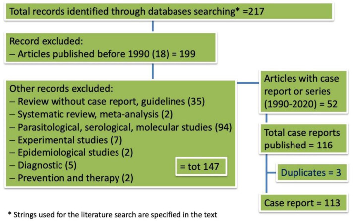
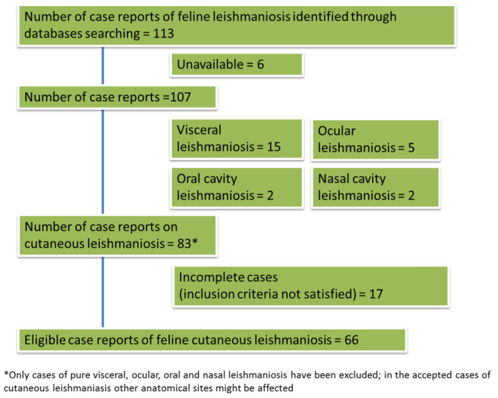
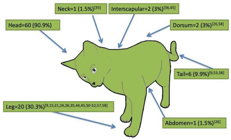
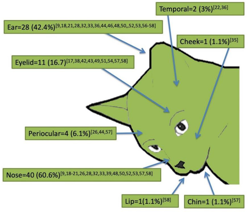
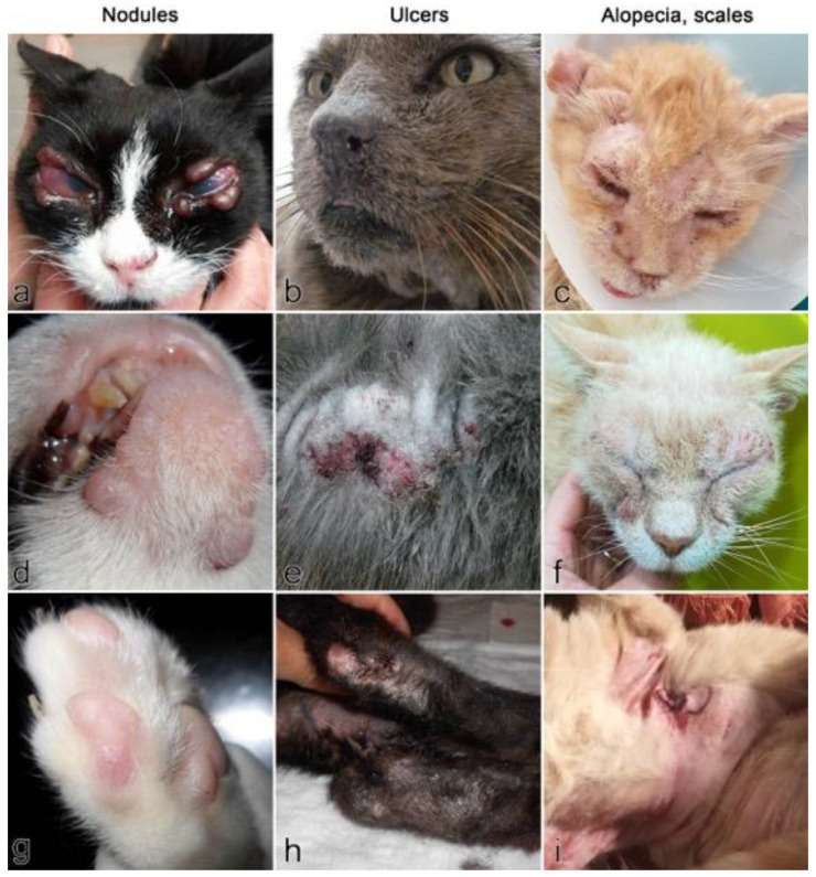
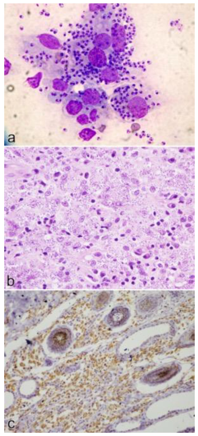
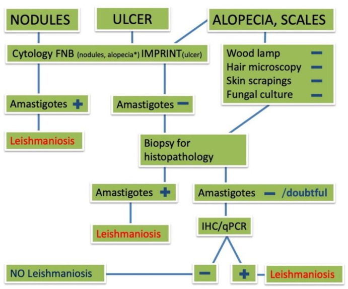
Feline leishmaniosis (FeL) is increasingly reported throughout the world and skin lesions predominate in the clinical picture. There are, however, few evidence-based data on cutaneous feline leishmaniosis and directions are strongly needed for a better management of the disease. In this study, we systematically reviewed what is currently known about the clinical dermatological presentation of FeL through analysis of the literature and, further, by adding unpublished cases managed by Italian veterinary dermatologists. Sixty-six feline cases of cutaneous leishmaniosis published in 33 articles between 1990 and 2020 met the inclusion criteria and were analyzed. Six unpublished cases of cutaneous FeL managed by Italian dermatologists were also reviewed. The majority of cases were reported from South America, followed by Europe and North America. Nodules were the most frequently reported clinical signs and the presence of Leishmania in lesioned skin was assessed mainly by cytology. A total of six Leishmania species have been identified as being responsible for skin lesions. Coinfections by FIV or FeLV were reported in 12.1% and 9.1% of the cases, respectively. Clinical data including treatment have been analyzed and discussed to provide directives for proper management of the disease for which cats may also serve as domestic reservoirs for human infections.

Keywords: Leishmania; cat; dermatology; feline leishmaniosis; skin lesions; systematic review.


Conflict of interest statement

The authors declare no conflict of interest.

Figures

Figure 1
Figure 1
Flowchart illustrating the results of database search to find cases of feline cutaneous leishmaniosis.
Figure 2
Figure 2
Flowchart illustrating the results of database search to find eligible cases of feline cutaneous leishmaniosis.
Figure 3
Figure 3
Anatomical distribution of lesions in the 66 reviewed cases of feline cutaneous leishmaniosis. References are reported as numbers between square brackets.
Figure 4
Figure 4
Anatomical distribution of lesions on the head in the 66 reviewed cases of feline cutaneous leishmaniosis. References are reported as numbers between square brackets.
Figure 5
Figure 5
Clinical signs in the six unpublished cases of feline cutaneous leishmaniosis. Types of lesions reported are skin nodules (a,d,g), ulcers (b,e,h), alopecia and scales (c,f,i). (a) Case no. 3: multiple alopecic and non-ulcerated nodules on both eyelids. (b) Case no. 5: small ulcer on the left nostril. (c) Case no. 6: symmetrical alopecic, scaly and crusted dermatitis on the face. (d) Case no. 4: multiple alopecic, non-ulcerated nodules on the inferior lip and chin. (e) Case no. 5: wide alopecic and ulcerated lesion on the dorsum. (f) Case no. 1: symmetrical alopecic, scaly and crusted dermatitis on the face. (g) Case no. 4: small alopecic, non-ulcerated nodule adjacent to the lateral digit. (h) Case no. 2: bilateral alopecic, scaly and ulcerated lesions of the metatarsal region. (i) Case no. 1: diffuse abdominal alopecia, erythema and scaly dermatitis.
Figure 6
Figure 6
Cytological, histological and immunohistochemical findings in cases of feline cutaneous leishmaniosis: (a) cytology of a nodule from case no. 4: large macrophages containing numerous amastigotes; (b) histology of a nodule from case no. 4: nodular to diffuse granulomatous dermatitis, with lymphocytes and numerous macrophages filled with amastigotes; (c) immunohistochemistry of skin lesion from case no. 2: many protozoal organisms are positive by using a hyperimmune canine serum.
Figure 7
Figure 7
Sequence step algorithm for the diagnosis of feline cutaneous leishmaniosis according to the type of skin lesion. FNB = fine needle biopsy; IHC = immunohistochemistry; qPCR = quantitative polymerase chain reaction.

References

    1. Gramiccia M. Recent advances in leishmaniosis in pet animals: Epidemiology, diagnostics and anti-vectorial prophylaxis. Vet. Parasitol. 2011;181:23–30. doi: 10.1016/j.vetpar.2011.04.019. - DOI - PubMed
    1. Solano-Gallego L., Koutinas A., Mirò G., Cardoso L., Pennisi M.G., Ferrer L., Bordeau P., Oliva G., Baneth G. Directions for the diagnosis, clinical staging, treatment and prevention of canine leishmaniosis. Vet. Parasitol. 2009;165:1–18. doi: 10.1016/j.vetpar.2009.05.022. - DOI - PubMed
    1. Roque A.L.R., Jansen A.M. Wild and synanthropic reservoirs of Leishmania species in the Americas. Int. J. Parasitol. Parasites Wildl. 2014;3:251–262. doi: 10.1016/j.ijppaw.2014.08.004. - DOI - PMC - PubMed
    1. Alemayehu B., Alemayehu M. Leishmaniasis: A Review on Parasite, Vector and Reservoir Host. Health Sci. J. 2017;11:519. doi: 10.21767/1791-809X.1000519. - DOI
    1. Kirkpatrick C.E., Farrell J.P., Goldschmidt M.H. Leishmania chagasi and L. donovani: Experimental Infections in Domestic Cats. Exp. Parasitol. 1984;58:125–131. doi: 10.1016/0014-4894(84)90027-4. - DOI - PubMed

LinkOut - more resources