. 2021 Apr 28;26(9):2566.
doi: 10.3390/molecules26092566.

Berberrubine Phosphate: A Selective Fluorescent Probe for Quadruplex DNA

Affiliations

Berberrubine Phosphate: A Selective Fluorescent Probe for Quadruplex DNA

Peter Jonas Wickhorst et al. Molecules. .

Abstract

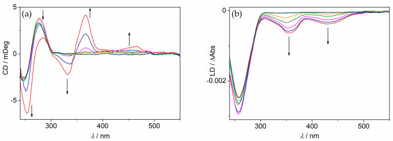
A phosphate-substituted, zwitterionic berberine derivative was synthesized and its binding properties with duplex DNA and G4-DNA were studied using photometric, fluorimetric and polarimetric titrations and thermal DNA denaturation experiments. The ligand binds with high affinity toward both DNA forms (Kb = 2-7 × 10⁵ M-1) and induces a slight stabilization of G4-DNA toward thermally induced unfolding, mostly pronounced for the telomeric quadruplex 22AG. The ligand likely binds by aggregation and intercalation with ct DNA and by terminal stacking with G4-DNA. Thus, this compound represents one of the rare examples of phosphate-substituted DNA binders. In an aqueous solution, the title compound has a very weak fluorescence intensity (Φfl < 0.01) that increases significantly upon binding to G4-DNA (Φfl = 0.01). In contrast, the association with duplex DNA was not accompanied by such a strong fluorescence light-up effect (Φfl < 0.01). These different fluorimetric responses upon binding to particular DNA forms are proposed to be caused by the different binding modes and may be used for the selective fluorimetric detection of G4-DNA.

Keywords: DNA ligand; G4-DNA; alkaloids; light-up probe; quadruplex DNA; zwitterion.


Conflict of interest statement

The authors declare no conflict of interest.

Figures

Scheme 1
Scheme 1
Synthesis of 9-O-Hydrogenphosphonatoberberrubin (2).
Figure 1
Figure 1
Emission spectra of 2 (c = 10 µM, with 0.25% v/v DMSO) in glycerol at 20 °C, 30 °C, 40 °C, 50 °C, 60 °C, and 80 °C; λex = 430 nm. The arrows indicate the changes in the emission intensity with increasing temperature. Inset: Plot of the relative emission intensity at 528 nm versus the temperature of the solution.
Figure 2
Figure 2
Photometric titration of 2 (c = 20 μM) with ct DNA (a) in a BPE buffer (cNa+ = 16 mM, pH 7.0, with 5% v/v DMSO) and with a2 (b) in a K-phosphate buffer (cK+ = 110 mM, pH 7.0, with 5% v/v DMSO). The arrows indicate the changes in absorption upon the addition of DNA. Inset: Plot of the absorption at 345 nm versus DNA concentration.
Figure 3
Figure 3
CD spectra (a) and LD spectra (b) of ct DNA (cDNA = 20 μM) in the absence and presence of 2 at LDR = 0 (black), 0.05 (cyan), 0.2 (orange), 0.5 (green), 1.0 (magenta), 1.5 (blue), and 2.0 (red) in a BPE buffer solution (10 mM, pH 7.0; with 5% v/v DMSO). The arrows indicate the changes in absorption with increasing LDR.
Figure 4
Figure 4
CD spectra of a2 (a, cDNA = 20 μM) and c-myc (b, cDNA = 20 μM) in the absence and presence of 2 at LDR = 0 (black), 0.05 (cyan), 0.2 (orange), 0.5 (green), 1.0 (magenta), 1.5 (blue), and 2.0 (red) in a K-phosphate buffer (cK+ = 110 mM, pH 7.0, with 5% v/v DMSO). The arrows indicate the changes in absorption with increasing LDR.
Figure 5
Figure 5
The proposed association of ligand 2 at the terminal quartet of G4-DNA c-myc and c-kit.
Figure 6
Figure 6
Fluorimetric titration of 2 (c = 20 μM, λex = 430 nm) with ct DNA (a) in a BPE buffer (cNa+ = 16 mM, pH 7.0, with 5% v/v DMSO) and with a2 (b) in a K-phosphate buffer (cK+ = 110 mM, pH 7.0, with 5% v/v DMSO); λex = 430 nm. The arrows indicate the changes in absorption upon addition of DNA. Inset: Plot of the relative fluorescence intensity versus cDNA; (c) pictures of the emission color of 2 in a K-phosphate buffer (i) and in the presence of ct DNA (ii) and 22AG (iii) (λex = 360 nm).

References

    1. Liu H., Lei Y. A critical review: Recent advances in “digital” biomolecule detection with single copy sensitivity. Biosens. Bioelectron. 2021;177:112901. doi: 10.1016/j.bios.2020.112901. - DOI - PMC - PubMed
    1. Yang X., Lovell J.F., Murthy N., Zhang Y. Topics in Medicinal Chemistry. Volume 34. Springer; Cham, Switzerland: 2020. pp. 33–53.
    1. Michel B.Y., Dziuba D., Benhida R., Demchenko A.P., Burger A. Probing of nucleic acid structures, dynamics, and interactions with environment-sensitive fluorescent labels. Front. Chem. 2020;8:112. doi: 10.3389/fchem.2020.00112. - DOI - PMC - PubMed
    1. Barnoy E.A., Popovtzer R., Fiixler D. Fluorescence for biological logic gates. J. Biophotonics. 2020;13:e202000158. doi: 10.1002/jbio.202000158. - DOI - PubMed
    1. Smith S.J., Nemr C.R., Kelley S.O. Chemistry-driven approaches for ultrasensitive nucleic acid detection. J. Am. Chem. Soc. 2017;139:1020–1028. doi: 10.1021/jacs.6b10850. - DOI - PubMed

LinkOut - more resources