Comparative Study
. 2021 Apr 28;22(9):4637.
doi: 10.3390/ijms22094637.

Species-Specific Regulation of TRPM2 by PI(4,5)P2 via the Membrane Interfacial Cavity

Affiliations
Comparative Study

Species-Specific Regulation of TRPM2 by PI(4,5)P2 via the Membrane Interfacial Cavity

Daniel Barth et al. Int J Mol Sci. .

Abstract

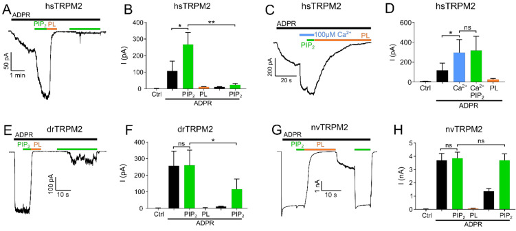
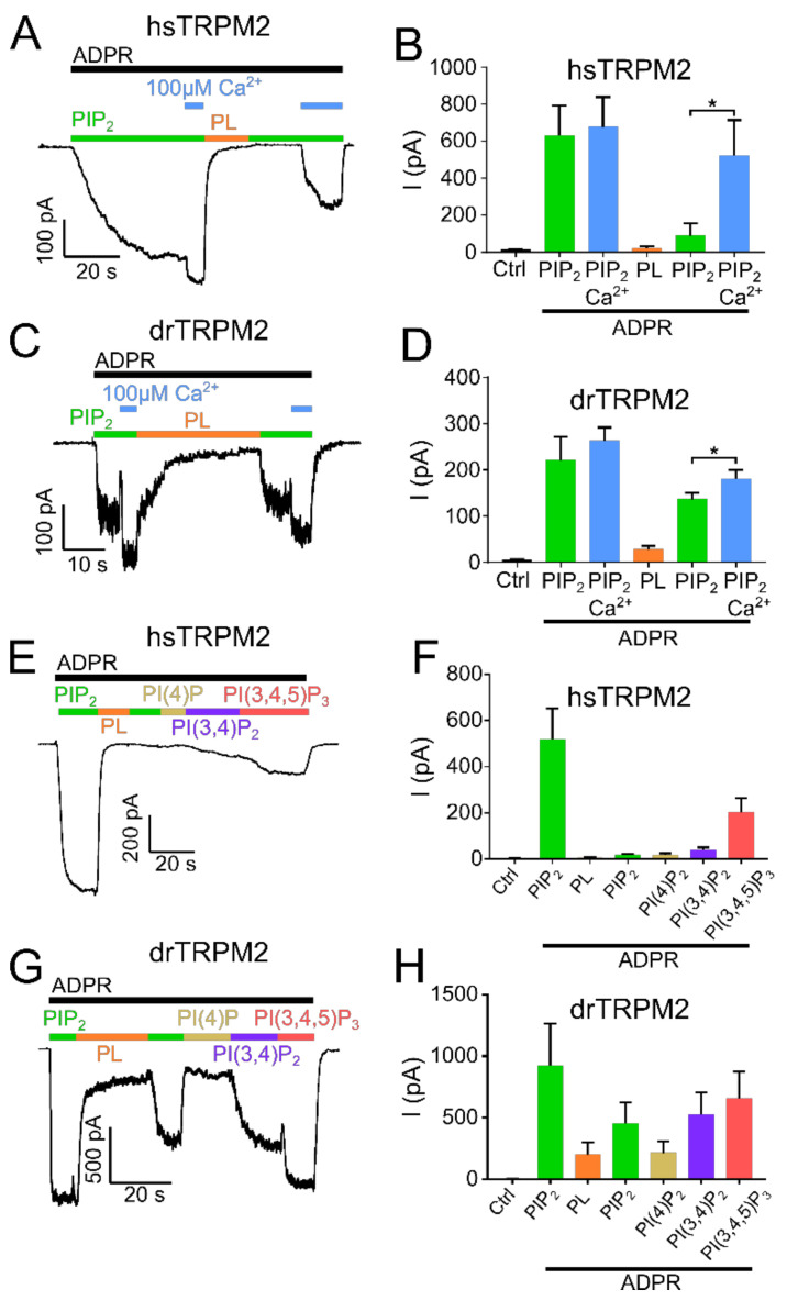
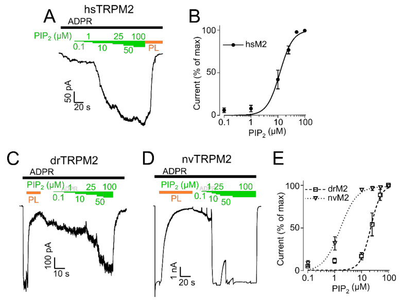
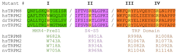
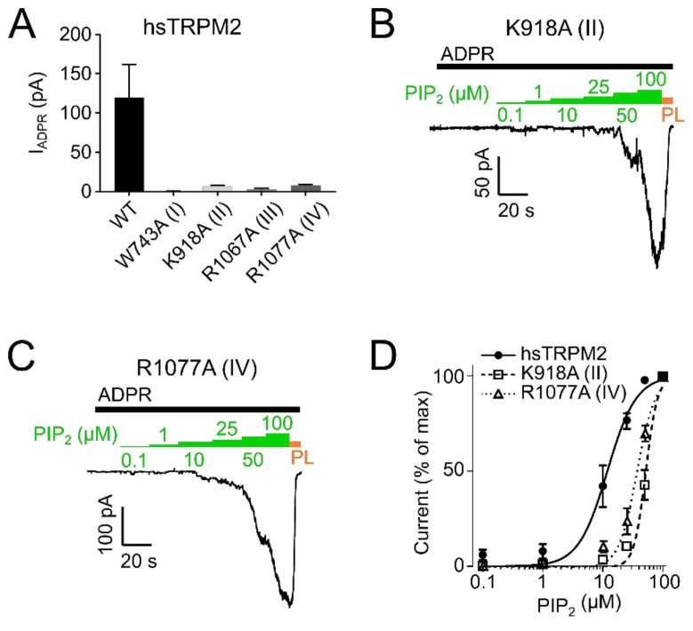
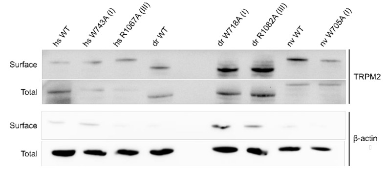
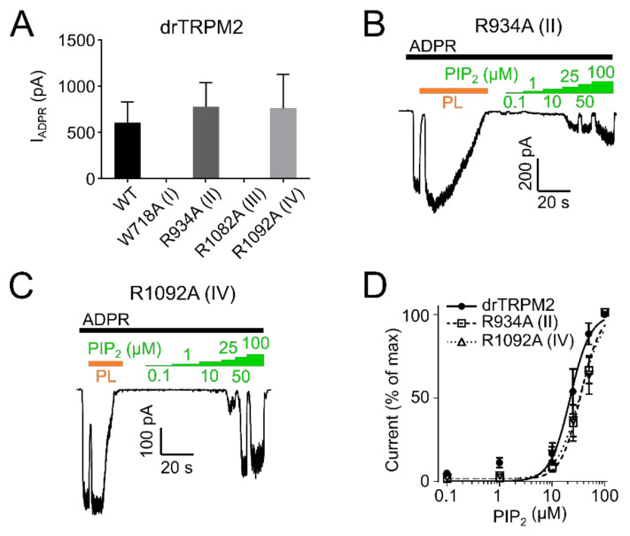
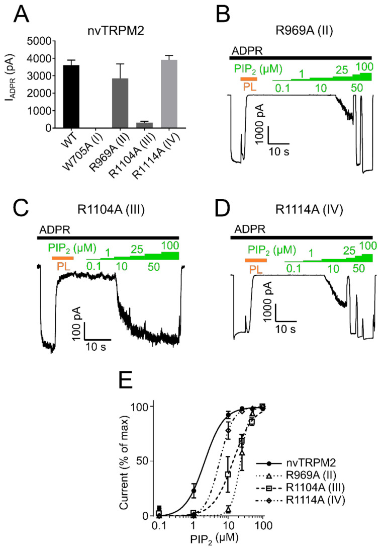
The human apoptosis channel TRPM2 is stimulated by intracellular ADR-ribose and calcium. Recent studies show pronounced species-specific activation mechanisms. Our aim was to analyse the functional effect of phosphatidylinositol 4,5-bisphosphate (PI(4,5)P2), commonly referred to as PIP2, on different TRPM2 orthologues. Moreover, we wished to identify the interaction site between TRPM2 and PIP2. We demonstrate a crucial role of PIP2, in the activation of TRPM2 orthologues of man, zebrafish, and sea anemone. Utilizing inside-out patch clamp recordings of HEK-293 cells transfected with TRPM2, differential effects of PIP2 that were dependent on the species variant became apparent. While depletion of PIP2 via polylysine uniformly caused complete inactivation of TRPM2, restoration of channel activity by artificial PIP2 differed widely. Human TRPM2 was the least sensitive species variant, making it the most susceptible one for regulation by changes in intramembranous PIP2 content. Furthermore, mutations of highly conserved positively charged amino acid residues in the membrane interfacial cavity reduced the PIP2 sensitivity in all three TRPM2 orthologues to varying degrees. We conclude that the membrane interfacial cavity acts as a uniform PIP2 binding site of TRPM2, facilitating channel activation in the presence of ADPR and Ca2+ in a species-specific manner.

Keywords: PIP2; TRPM2; patch clamp; phosphoinositides; phospholipid.


Conflict of interest statement

The authors declare no conflict of interest.

Figures

Figure 1
Figure 1
PIP2 is a necessary cofactor for TRPM2 activation. Inside-out patch clamp recordings of HEK-293 cells expressing TRPM2 at −60 mV and 1 µM Ca2+ unless stated otherwise. Currents were activated by cytosolic exposure to ADPR (black bar). (A) Representative current trace of hsTRPM2 activated by cytosolic exposure to 300 µM ADPR (black bar) and 25 µM PIP2 (green bar) and blocked by 15 µg/mL polylysine (PL, orange bar). (B) Summary of the effects of ADPR, PIP2, and polylysine on hsTRPM2 currents. Statistical analysis reveals a significant current increase via PIP2 after the initial ADPR induced current plateaued. The current was abolished upon exposure to polylysine and unable to recover via a second PIP2 application. Control (Ctrl) represents standard bath solution in absence of ADPR. (C,D) Representative current trace and statistics of the PIP2 effect following elevated intracellular Ca2+ concentrations (100 µM) for hsTRPM2. Application of 100 µM Ca2+ causes a strong current increase in hsTRPM2 which is only slightly further increased upon external PIP2 addition. Current traces of drTRPM2 (E) and nvTRPM2 (G) exposed to 100 µM ADPR, 25 µM PIP2 and 15 µg/mL polylysine. Statistical analysis shows no significant difference for drTRPM2 (F) and nvTRPM2 (H) between the current observed during exposure to ADPR and the additional application of PIP2. (F) Fractional reactivation of drTRPM2 currents by second PIP2 application after the current was abolished via polylysine while nvTRPM2 (H) fully recovered. Data are presented as mean ± SEM analysed via one-way ANOVA with Dunn’s multiple comparison test; n = 3–8; * p < 0.05, ** p < 0.01, ns = not significant.
Figure 2
Figure 2
Influence of high calcium and PIP2 derivatives on TRPM2 current restoration. Recordings of HEK-293 cells expressing TRPM2 at −60 mV in the inside-out patch clamp configuration. Currents were activated by cytosolic exposure to ADPR (black bar) and PIP2 (25 µM) (green bar). Representative current trace and statistics of hsTRPM2 (A,B) and drTRPM2 (C,D) at high Ca2+ concentrations (100 µM). High Ca2+ in presence of PIP2 and ADPR (blue bar) causes current restoration after the current was inhibited via 15 µg/mL polylysine (PL, orange bar). Representative recordings and summary of current statistics of the effect of PI derivatives on hsTRPM2 (E,F) and drTRPM2 (G,H) currents using 1 µM cytosolic Ca2+. Statistical analysis was performed via paired Student’s t-test and data presented as mean ± SEM; n = 4–7; * p < 0.05.
Figure 3
Figure 3
PIP2 sensitivity of TRPM2. PIP2 dose-response recordings of inside-out patches of HEK-293 cells expressing hsTRPM2 (A), drTRPM2 (C), and nvTRPM2 (D). ADPR (black bar) was used to initiate TRPM2 currents followed by increasing concentrations of PIP2 (green bars). Polylysine (PL, orange bar) was utilized to scavenge PIP2. Summary of the PIP2 dose-response recordings calculated as a percentage of the maximum current obtained at 100 µM PIP2 (hsTRPM2 (B), drTRPM2, and nvTRPM2 (E)). Data were analysed from five to six independent experiments.
Figure 4
Figure 4
Sequence alignment of TRPM2 and TRPM8. Sequence alignment of the membrane interfacial cavity (formed by: Pre-S1 domain and MHR4 from the adjacent subunit (green), junction between S4 and S5 (purple) and TRP domain (orange)) of hs-, dr- and nvTRPM2 and hsTRPM8. Target positions (yellow) within MHR4-PreS1 labelled mutant I (hsW743, drW718, and nvW705), S4–S5 linker labelled mutant II (hsK918, drR934, and nvR969) and TRP domain separated in mutant III (hsR1067, drR1082, nvR1104) and mutant IV (hsR1077, drR1092, nvR1114).
Figure 5
Figure 5
PIP2 sensitivity of hsTRPM2 membrane interfacial cavity mutant channels. (A) Current amplitudes of wild-type and mutant hsTRPM2 channels in response to 300 µM ADPR. Representative current trace of K918A (II) (B) and R1077A (IV) (C) exposed to increasing PIP2 (green bar) concentrations in the presence of 300 µM ADPR (black bar). 15 µg/mL polylysine (PL, orange bar) was used to block PIP2 mediated currents. (D) Data summary of PIP2 dose-response experiments showing a clear right shift of mutants K918A (II) and R1077A (IV) compared with the wild-type. Data presented as mean ± SEM; n = 5.
Figure 6
Figure 6
Biotinylation assay of TRPM2 expressed in HEK-293 cells. Representative surface and total expression of hs-, dr- and nvTRPM2 WT and point mutations via biotinylation. β-actin was used as the loading control.
Figure 7
Figure 7
PIP2 sensitivity of drTRPM2 membrane interfacial cavity mutant channels. (A) ADPR (100 µM, black bar) induced currents of wild-type and mutant drTRPM2 channels. Current trace of R934A (II) (B) and R1092A (IV) (C) perfused with increasing concentrations of PIP2 (green bars). Polylysine (15 µg/mL, PL, orange bar) was used to scavenge PIP2 since even in the absence of external PIP2 the channels are already saturated with natural membrane PIP2. (D) Summary of the presented data showing a right shift in PIP2 sensitivity of mutants R934A (II) and R1092A (IV). Data represented as mean ± SEM; n = 5–6.
Figure 8
Figure 8
PIP2 sensitivity of nvTRPM2 membrane interfacial cavity mutant channels. (A) Current amplitudes induced by ADPR (100 µM, black bar) of wild-type and mutant nvTRPM2 channels. PIP2 (green bars) dose-response recordings of nvTRPM2 R969A (II) (B), R1104A (III) (C) and R1114A (IV) (D) in presence of 100 µM ADPR (black bar). Since maximum currents were already observed in absence of external PIP2, natural PIP2 was scavenged via polylysine (15 µg/mL, PL, orange bar) and subsequently restored via increasing PIP2 concentrations. (E) Data summary of PIP2 sensitivity of mutants R969A (II), R1104A (III), and R1114A (IV) showing dominant right shift compared with wild-type nvTRPM2. Data represented as mean ± SEM; n = 5–6.

References

    1. Kuhn F.J.P. Structure-function relationship of TRPM2: Recent advances, contradictions, and open questions. Int. J. Mol. Sci. 2020;21:6481. doi: 10.3390/ijms21186481. - DOI - PMC - PubMed
    1. Sano Y., Inamura K., Miyake A., Mochizuki S., Yokoi H., Matsushime H., Furuichi K. Immunocyte Ca2+ influx system mediated by LTRPC2. Science. 2001;293:1327–1330. doi: 10.1126/science.1062473. - DOI - PubMed
    1. Perraud A.L., Fleig A., Dunn C.A., Bagley L.A., Launay P., Schmitz C., Stokes A.J., Zhu Q., Bessman M.J., Penner R., et al. ADP-ribose gating of the calcium-permeable LTRPC2 channel revealed by Nudix motif homology. Nature. 2001;411:595–599. doi: 10.1038/35079100. - DOI - PubMed
    1. Csanady L., Torocsik B. Four Ca2+ ions activate TRPM2 channels by binding in deep crevices near the pore but intracellularly of the gate. J. Gen. Physiol. 2009;133:189–203. doi: 10.1085/jgp.200810109. - DOI - PMC - PubMed
    1. McHugh D., Flemming R., Xu S.Z., Perraud A.L., Beech D.J. Critical intracellular Ca2+ dependence of transient receptor potential melastatin 2 (TRPM2) cation channel activation. J. Biol. Chem. 2003;278:11002–11006. doi: 10.1074/jbc.M210810200. - DOI - PubMed

Publication types

Substances