Review
. 2021 Apr 28;9(5):483.
doi: 10.3390/biomedicines9050483.

Optoacoustic Imaging in Inflammation

Affiliations
Review

Optoacoustic Imaging in Inflammation

Adrian P Regensburger et al. Biomedicines. .

Abstract

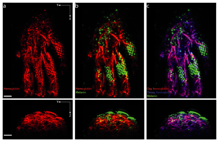
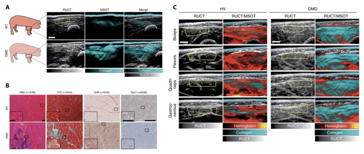
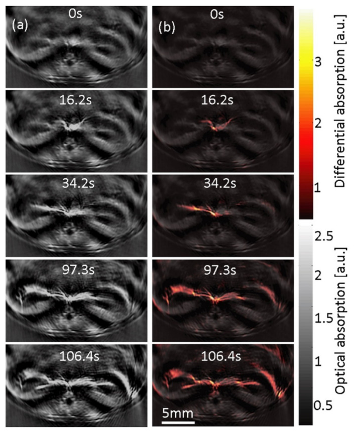
Optoacoustic or photoacoustic imaging (OAI/PAI) is a technology which enables non-invasive visualization of laser-illuminated tissue by the detection of acoustic signals. The combination of "light in" and "sound out" offers unprecedented scalability with a high penetration depth and resolution. The wide range of biomedical applications makes this technology a versatile tool for preclinical and clinical research. Particularly when imaging inflammation, the technology offers advantages over current clinical methods to diagnose, stage, and monitor physiological and pathophysiological processes. This review discusses the clinical perspective of using OAI in the context of imaging inflammation as well as in current and emerging translational applications.

Keywords: MSOT; PAI; RSOM; acute inflammation; chronic inflammation; imaging inflammation; molecular imaging; optoacoustics; photoacoustics.


Conflict of interest statement

A.P.R., M.J.W., and F.K. are co-inventors, together with iThera Medical GmbH, Munich, Germany, on an EU patent application (EP 19 163 304.9) relating to a device and a method for analyzing optoacoustic data, an optoacoustic system, and a computer program. A.P.R. and F.K. received travel support from iThera Medical GmbH, Munich, Germany. A.P.R. reports lecture fees from Sanofi-Aventis Deutschland GmbH, Frankfurt, Germany. F.K. reports lecture fees from Siemens Healthcare GmbH, Erlangen, Germany outside the submitted work. All other authors declare no competing interests.

Figures

Figure 4
Figure 4
Multispectral optoacoustic tomography (MSOT) of intestinal inflammation using hemoglobin as a surrogate for disease activity. The technique enables hybrid imaging together with B-mode ultrasound imaging (RUCT). (A) MSOT imaging of the liver in humans enabling the generation of signals for different subcellular compounds; (B) S = serosa, M = muscularis mucosa, Sm = submucosa, Mu = mucosa, L = lumen, RUCT = reflectance ultrasound computed tomography. Figure (A) reproduced and modified from [131]. (https://creativecommons.org/licenses/by/4.0/ (accessed on 9 April 2021)).
Figure 1
Figure 1
Absorption coefficients (µa in cm−1) versus wavelength (in nm) for different optoacoustic imaging molecules and tissue. Spectra were derived from existing data as indicated: melanin (https://omlc.org/spectra/melanin/mua.html as derived from [15,16,17,18]), oxy- (HbO2) and deoxyhemoglobin (HbR) (https://omlc.org/spectra/hemoglobin/summary.html), (bulk) lipid (https://omlc.org/spectra/fat/fat.txt as derived from [19]), water (https://omlc.org/spectra/water/data/hale73.txt derived from [20]), aorta tissue (https://omlc.org/spectra/aorta/oraevsky_a.txt derived from [21]), and collagen (extracted from [22]). All databases accessed on 9 April 2021.
Figure 2
Figure 2
Inflammation is a multistep process triggered by a variety of causes and agents. This results in acute and/or chronic inflammation, which leads to host response in terms of cell recruitment, adaptions of the vasculature, and changes of the tissue composition and extracellular matrix. All of these aspects pose possible targets for opto-/photoacoustic imaging. Figure created with BioRender.com.
Figure 3
Figure 3
Raster-scanning optoacoustic mesoscopy enables a precise visualization of microvasculature. Depending on the detection frequencies, larger (11–33 MHz, in red) or smaller (33–99 MHz, in green) vessels can be resolved. White square indicates magnified area. Scale bar indicates 1 mm.
Figure 5
Figure 5
Raster-scanning optoacoustic mesoscopy (RSOM) of murine paw vasculature and the potential imaging readouts for inflammation. (a) Shows the hemoglobin signal representing the vascular network, which can be used to identify changes in blood volume; (b) multi-wavelength illumination enables the separation of melanin and hemoglobin signals; (c) further unmixing enables the visualization of oxygenation status of hemoglobin. Scale bar indicates 1mm.
Figure 6
Figure 6
Optoacoustic imaging (OAI) of muscle degeneration in Duchenne muscular dystrophy. (A) Shows signals derived from wild-type (WT) and Duchenne muscular dystrophy (DMD) transgenic pigs; (B) demonstrates ex vivo tissue changes and expansion of extracellular matrix and collagens in diseased tissues as a possible correlate of OAI signals; (C) signals derived from healthy volunteers (HV) and pediatric patients with DMD at different anatomical positions. RUCT and RUCT/MSOT merged images shown. RUCT = reflectance ultrasound computed tomography, MSOT = multispectral optoacoustic tomography. Signals unmixed for hemoglobin and collagen. Figure modified with permission from [94]. This image is not published under the terms of the CC-BY license. For permission to reuse, please see [72].
Figure 7
Figure 7
Tomographic optoacoustic imaging of indocyanine perfusion kinetics in murine kidneys. (a) Cross-sectional optoacoustic images over time of mouse kidneys at 800 nm after ICG injection; (b) the absorption difference with the single-wavelength image before injection to show increased ICG perfusion over time. Adapted with permission from [95]. This image is not published under the terms of the CC-BY license. © The Optical Society.

References

    1. Wilting J., Becker J., Buttler K., Weich H.A. Lymphatics and inflammation. Curr. Med. Chem. 2009;16:4581–4592. doi: 10.2174/092986709789760751. - DOI - PubMed
    1. Gupta S.C., Kunnumakkara A.B., Aggarwal S., Aggarwal B.B. Inflammation, a Double-Edge Sword for Cancer and Other Age-Related Diseases. Front. Immunol. 2018;9:2160. doi: 10.3389/fimmu.2018.02160. - DOI - PMC - PubMed
    1. Karin M., Clevers H. Reparative inflammation takes charge of tissue regeneration. Nature. 2016;529:307–315. doi: 10.1038/nature17039. - DOI - PMC - PubMed
    1. Hammoud D.A. Molecular Imaging of Inflammation: Current Status. J. Nucl. Med. 2016;57:1161–1165. doi: 10.2967/jnumed.115.161182. - DOI - PMC - PubMed
    1. Willmann J.K., van Bruggen N., Dinkelborg L.M., Gambhir S.S. Molecular imaging in drug development. Nat. Rev. Drug Discov. 2008;7:591–607. doi: 10.1038/nrd2290. - DOI - PubMed

LinkOut - more resources