. 2021 Apr 28;9(5):482.
doi: 10.3390/biomedicines9050482.

Celastrol and Triptolide Suppress Stemness in Triple Negative Breast Cancer: Notch as a Therapeutic Target for Stem Cells

Affiliations

Celastrol and Triptolide Suppress Stemness in Triple Negative Breast Cancer: Notch as a Therapeutic Target for Stem Cells

Prabhu Ramamoorthy et al. Biomedicines. .

Abstract

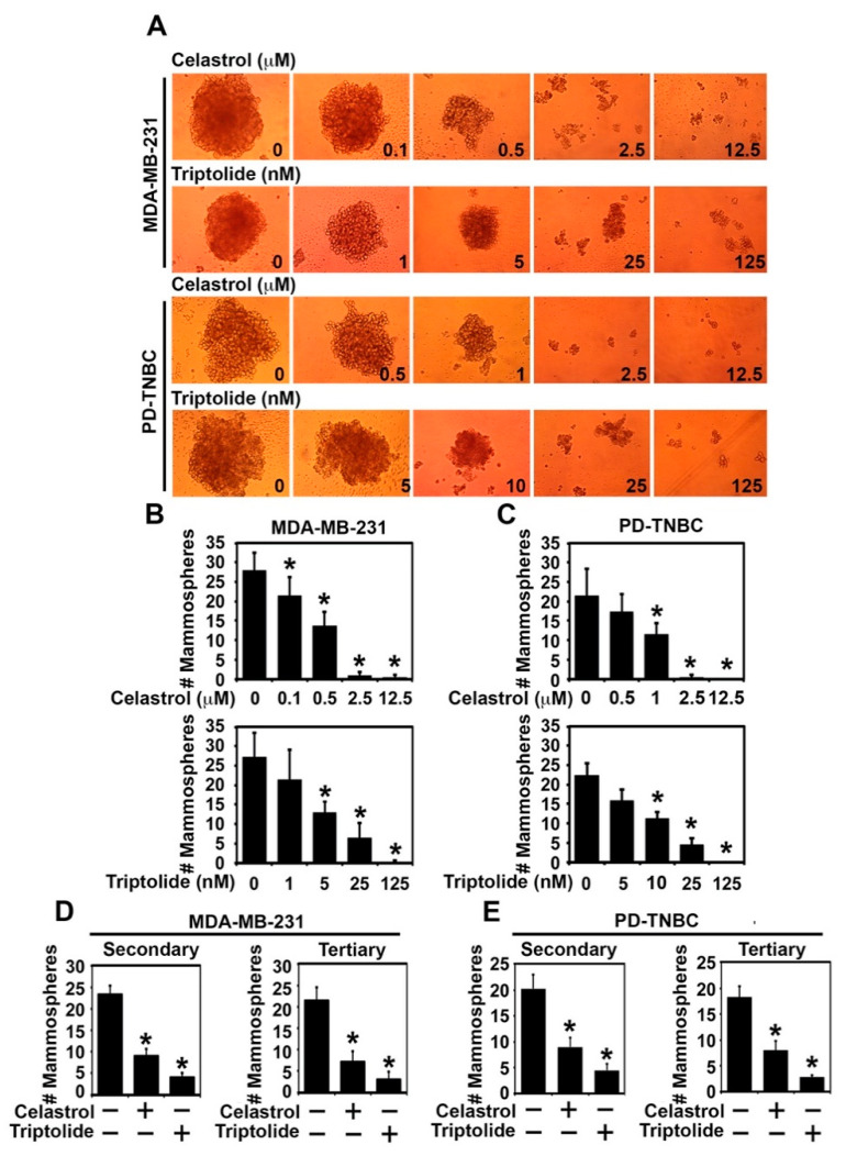
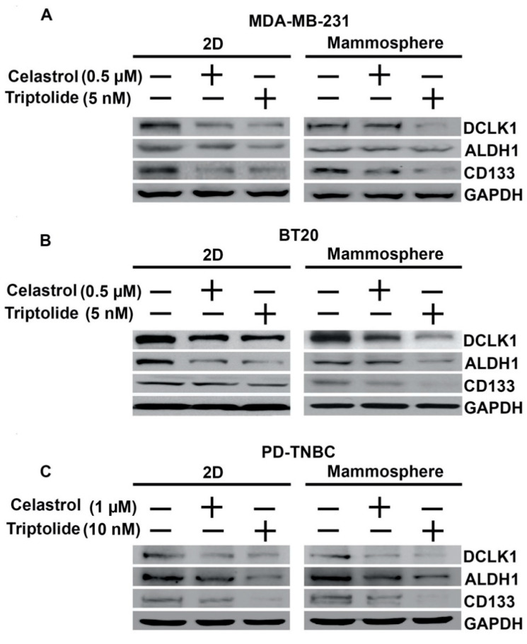
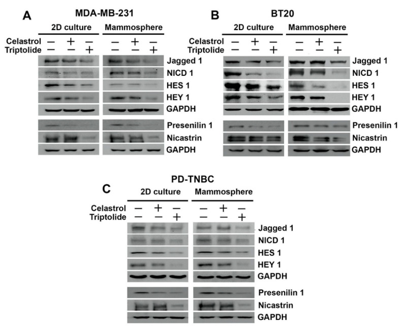
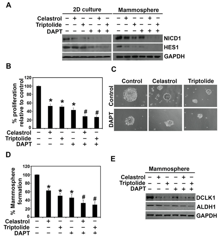
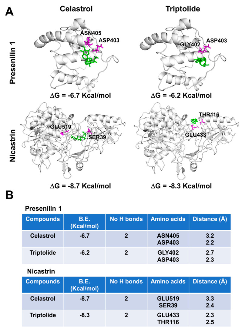
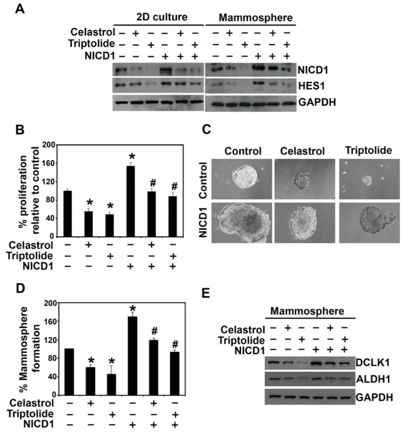
Triple negative breast cancer (TNBC) is observed in ~15% of breast cancers and results in poor survival and increased distant metastases. Within the tumor are present a small portion of cancer stem cells that drive tumorigenesis and metastasis. In this study, we aimed to elucidate whether the two natural compounds, celastrol and triptolide, inhibit stemness in TNBC. MDA-MB-231, BT20, and a patient-derived primary cells (PD-TNBC) were used in the study. Mammosphere assay was performed to assess the stemness. Both celastrol and triptolide treatment suppressed mammosphere formation. Furthermore, the compound suppressed expression of cancer stem cell marker proteins DCLK1, ALDH1, and CD133. Notch signaling plays a critical role in stem cells renewal. Both celastrol or triptolide reduced Notch -1 activation and expression of its downstream target proteins HES-1 and HEY-1. However, when NICD 1 was ectopically overexpressed in the cells, it partially rescued proliferation and mammosphere formation of the cells, supporting the role of notch signaling. Together, these data demonstrate that targeting stem cells and the notch signaling pathway may be an effective strategy for curtailing TNBC progression.

Keywords: ALDH1; DCLK1; mammospheres; nicastrin; presenilin; γ-secretase.


Conflict of interest statement

The authors have no financial disclosure.

Figures

Figure 1
Figure 1
Celastrol and triptolide reduce mammosphere growth and number in TNBC cells. A and B) MDA-MB-231 and PD-TNBC cells were treated with celastrol (0–12.5 µM) and triptolide (0–125 nM). (A) Representative images show reduction in mammosphere size. (B,C) Total number (#) of primary mammospheres was reduced in a dose-dependent manner following celastrol and triptolide treatment when compare to control. * p < 0.05 when compared with control. (D,E) Primary mammospheres of MDA-MB-231, PD-TNBC cells treated with celastrol (0.5 µM) and triptolide (5 nM) were first treated with celastrol (1µm) and triptolide (10 nm), and subsequently used to grow secondary and tertiary mammospheres without drug treatment. * p < 0.05 when compared with control.
Figure 2
Figure 2
Celastrol and Triptolide attenuate expression of stem cell markers in TNBC cells. The two compounds inhibit cancer stem cells marker, DCLK1, ALDH1, and CD133 protein expression as compared to control, untreated cells in both 2D culture (left panel) and mammosphere cultures (right panel). (A) MDA-MB-231, (B) BT20, and (C) PD-TNBC.
Figure 3
Figure 3
HSP90 inhibitors affect notch signaling in TNBC cells. Celastrol and triptolide inhibited notch signaling by decreasing the expression level of Notch ligand, Jagged-1, NICD 1, and Notch downstream target proteins HES1 and HEY1 in TNBC cells. Left panel shows blots from cells grown in 2D cell and right panel shows those from mammospheres. (A) MDA-MB-231 cells, (B) BT20 cells, and (C) PD-TNBC cells.
Figure 4
Figure 4
γ-secretase inhibitor, DAPT, altered the celastrol and triptolide-induced suppression of notch signaling in TNBC cells. (A) Western blot analysis of BT20 cells treated with DAPT, celastrol, and triptolide for 48 h. NICD1 and HES1 protein expression were suppressed in celastrol and triptolide treatment and further decreased in combination with DAPT. (B) Cell proliferation was significantly inhibited following treatment with the compounds and further reduced in combination of DAPT. Each value represents mean ± SD of four separate estimations, n = 3. * p < 0.05 when compared with control; # p < 0.05 when compared with DAPT control. (C,D) DAPT treatment alone reduces the mammosphere formation when compared to the control, and a combination with celastrol and triptolide shows further reduction in mammosphere growth. Each value represents mean ± SD of four separate estimations, n = 3. * p < 0.05 when compared with control; # p < 0.05 when compared with DAPT control. (E) Expression of stem cell marker proteins DCLK1 and ALDH1 is reduced following DAPT treatment. Combination treatment with celastrol or triptolide further decreases the expression of these proteins in mammospheres.
Figure 5
Figure 5
Celastrol and Triptolide bind to γ-secretase proteins presenilin 1 and nicastrin. (A) Binding of Celastrol and Triptolide in the protein cavity of presenilin 1 and nicastrin were assessed by molecular docking technique using Autodock vina software program. Celastrol and Triptolide bind to presenilin 1 and nicastrin with the binding energy (B.E.) of −6.7 and −7.2 Kcal/mol respectively, as well as bind to nicastrin with B.E. of −8.7 and −8.3 Kcal/mol respectively. Cartoon models are represented in the figure. (B) The docking results and consensus scores of Celastrol and Triptolide binding to presenilin 1 and nicastrin are summarized.
Figure 6
Figure 6
Overexpression of NICD1 partially protects from celastrol and triptolide treatment in BT20 cells. (A) NICD1 overexpression rescues cells from celastrol and triptolide-mediated suppression of NICD1 and HES1 expression. The cells were treated with the compounds for 48 h in cells overexpressing NICD1, and cell lysates were analyzed by western blotting. Increased protein expression of NICD1 and HES-1 was observed in the NICD-expressing cells when compared with vector controls. Celastrol and triptolide reduced the expression of NICD1 and HESI., but this was partially restored in NICD1 overexpressing cells. (B) NICD1-overexpressing BT20 cells overcome the suppression of cell growth in celastrol and triptolide. Each value represents mean ± SD of four separate estimations, n = 3. * p < 0.05 when compared with vector control; # p < 0.05 when compared with NICD1 overexpression control. (C,D) Mammosphere cultures show partial protection from celastrol and triptolide-induced spheroid growth in NICD1 overexpressing cells. Each value represents mean ± SD of four separate estimations, n = 3. * p < 0.05 when compared with vector control; # p < 0.05 when compared with NICD1 overexpression control. (E) DCLK1 and ALDH1 protein expression were decreased in celastrol and triptolide treatment but restored in NICD1 overexpressing cells after treatment.

References

    1. Siegel R.L., Miller K.D., Jemal A. Cancer Statistics, 2017. CA Cancer J. Clin. 2017;67:7–30. doi: 10.3322/caac.21387. - DOI - PubMed
    1. Rouzier R., Perou C.M., Symmans W.F., Ibrahim N., Cristofanilli M., Anderson K., Hess K.R., Stec J., Ayers M., Wagner P., et al. Breast cancer molecular subtypes respond differently to preoperative chemotherapy. Clin. Cancer Res. 2005;11:5678–5685. doi: 10.1158/1078-0432.CCR-04-2421. - DOI - PubMed
    1. Exman P., Tolaney S.M. HER2-positive metastatic breast cancer: A comprehensive review. Clin. Adv. Hematol. Oncol. 2021;19:40–50. - PubMed
    1. Fong P.C., Boss D.S., Yap T.A., Tutt A., Wu P., Mergui-Roelvink M., Mortimer P., Swaisland H., Lau A., O’Connor M.J., et al. Inhibition of poly(ADP-ribose) polymerase in tumors from BRCA mutation carriers. N. Engl. J. Med. 2009;361:123–134. doi: 10.1056/NEJMoa0900212. - DOI - PubMed
    1. Ramalho-Santos M., Yoon S., Matsuzaki Y., Mulligan R.C., Melton D.A. "Stemness": Transcriptional profiling of embryonic and adult stem cells. Science. 2002;298:597–600. doi: 10.1126/science.1072530. - DOI - PubMed

LinkOut - more resources