. 2021 Apr 28;13(5):788.
doi: 10.3390/v13050788.

AAV-HDV: An Attractive Platform for the In Vivo Study of HDV Biology and the Mechanism of Disease Pathogenesis

Affiliations

AAV-HDV: An Attractive Platform for the In Vivo Study of HDV Biology and the Mechanism of Disease Pathogenesis

Sheila Maestro et al. Viruses. .

Abstract

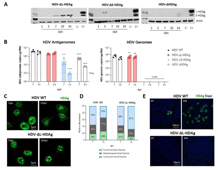
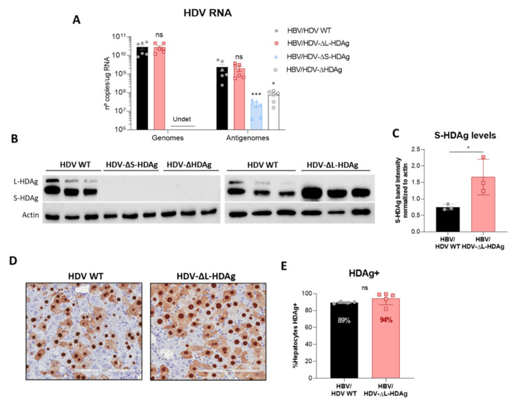
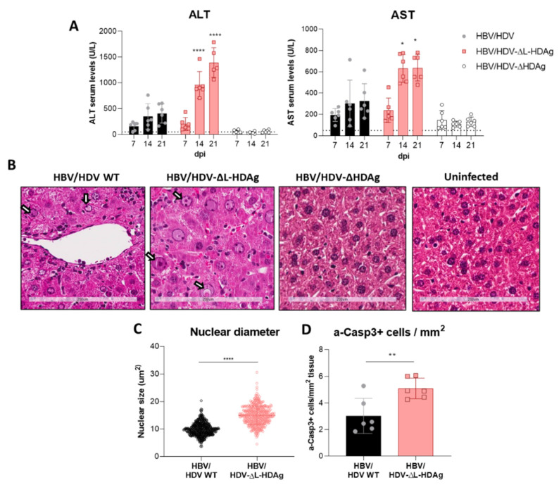
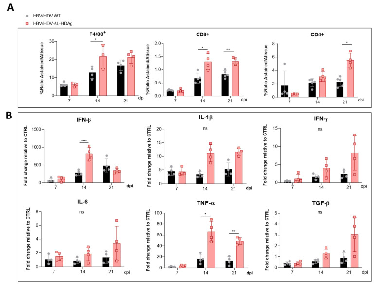
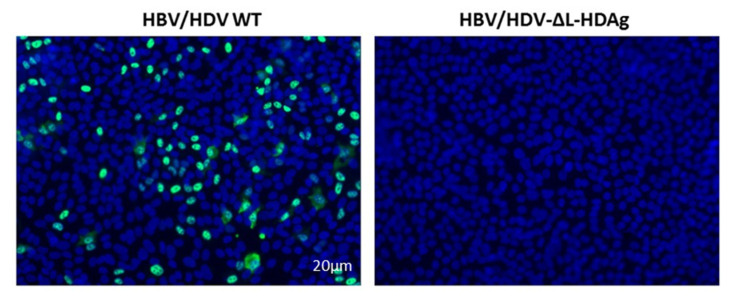
Hepatitis delta virus (HDV) infection causes the most severe form of viral hepatitis, but little is known about the molecular mechanisms involved. We have recently developed an HDV mouse model based on the delivery of HDV replication-competent genomes using adeno-associated vectors (AAV), which developed a liver pathology very similar to the human disease and allowed us to perform mechanistic studies. We have generated different AAV-HDV mutants to eliminate the expression of HDV antigens (HDAgs), and we have characterized them both in vitro and in vivo. We confirmed that S-HDAg is essential for HDV replication and cannot be replaced by L-HDAg or host cellular proteins, and that L-HDAg is essential to produce the HDV infectious particle and inhibits its replication. We have also found that lack of L-HDAg resulted in the increase of S-HDAg expression levels and the exacerbation of liver damage, which was associated with an increment in liver inflammation but did not require T cells. Interestingly, early expression of L-HDAg significantly ameliorated the liver damage induced by the mutant expressing only S-HDAg. In summary, the use of AAV-HDV represents a very attractive platform to interrogate in vivo the role of viral components in the HDV life cycle and to better understand the mechanism of HDV-induced liver pathology.

Keywords: AAV; HDAg; HDV; liver damage; mouse model.


Conflict of interest statement

The authors declare no conflict of interest in relation with the work presented in this manuscript.

Figures

Figure A1
Figure A1
The levels of shuttle plasmid containing the HDV antigenome decrease over time coinciding with cell division. Total DNA was extracted from HepG2 and Huh-7 cells and HDV DNA copies were quantified by qPCR.
Figure A2
Figure A2
Huh-7 cells were transfected with HDV WT or HDV-∆L-HDAg plasmid and they were harvested at 7 and 14 dpt. Cellular extracts were fractionated to determine the subcellular localization of S-HDAg and L-HDAg. The purity of each subcellular fractions was determined by analysing the expression of GAPDH (control of cytosolic-enriched fraction), EGFR (control of membrane fraction), and Histone2A (control of nuclear-enriched fraction).
Figure A3
Figure A3
HDV genome copies increase with time in C57BL/6 mice after AAV-HDV WT or AAV-HDV-ΔL-HDAg injection while the AAV genomes drop during the experiment. (A) HDV RNA was analyzed in livers of mice injected with AAV-HDV WT and AAV-HDV-ΔL-HDAg by RT-qPCR. (B) Total DNA was extracted from infected livers and AAV-HDV genomes were quantified by qPCR. Statistical analysis by two-way ANOVA showed significant differences in HDV RNA at 14 dpi. * p < 0.05.
Figure 1
Figure 1
The HDV life cycle is supported in Huh-7 cells upon transfecting HDV-encoding plasmid but not in HepG2 cells. (A) Schematic representation of the experimental layout. HepG2 or Huh-7 cells were seeded and transfected with equal amounts of the plasmid encoding the HDV antigenome. For RNA and protein analysis, cells were collected at 1-, 3-, 7-, 10- and 14-days post-transfection (dpt), and cells were split 1:3 at 7-dpt. (B) Total RNA was extracted from cells and HDV antigenome and genome levels were assessed by RT-qPCR. (C) Western Blot analysis of HepG2 and Huh-7 cell lysates was performed to detect S-HDAg and L-HDAg. Positive control (C+): Huh-7 cells transfected with plasmids expressing S-HDAg and L-HDAg antigens and collected at 3 dpt. Negative control (C-): non transfected Huh-7 or HepG2 cells. (D) IFN-β (left) and MxA (right) expression levels were quantified by RT-qPCR and normalized using GAPDH as housekeeping gene. Statistical analysis using Mann–Whitney test revealed differences between the two cell lines (*** p < 0.001, **** p < 0.0001).
Figure 2
Figure 2
HDV mutants affecting S-HDAg and L-HDAg expression were generated and characterized in Huh-7 cells. (A) HDAg expression was assessed in Huh-7 cell lysates by Western Blot at 1-, 3-, 7-, 10- and 14-dpt. (B) HDV genomes were produced after transfection with the plasmids that express S-HDAg: HDV WT and HDV-ΔL-HDAg. (ns: no significant, *** p < 0.001, **** p < 0.0001). (C,D) HDAg localization was examined by immunofluorescence (scale bar: 5 μm) and by cell fractionation. The percentage of HDAgs present in the cytosolic-, the membrane- and the nuclear-enriched fractions was determined at 7- and 14-dpt. (E) Huh-7.5.1-hNTCP cells were infected with supernatants from HBV/HDV WT, and HBV/HDV-ΔL-HDAg co-transfected cells collected at 7- and 14-dpt. At 7 days post-infection (dpi), cells were fixed and immune stained with human serum for HDAg detection. Scale bar: 10 μm.
Figure 3
Figure 3
Administration of AAV-HDV mutants lacking the expression of S-HDAg resulted in a loss of HDV replication, while the absence of L-HDAg enhances S-HDAg production. Eight-week-old C57BL/6 WT mice received (n = 6 mice/group) 5 × 10¹⁰ genome copies of AAV-HDV, AAV-HDV-ΔL-HDAg, AAV-HDV-ΔS-HDAg and AAV-HDV-ΔHDAg in combination with 5 × 10¹⁰ genome copies of AAV-HBV. (A) HDV genomes and antigenomes were analyzed in the livers of infected mice at 21 dpi (significant differences were determined by Kruskal-Wallis test and Dunn’s post-test). (Undet: undetectable; ns: no significant, * p < 0.05, *** p < 0.001). (B) Western blot analysis of liver samples revealed the absence of HDAgs in mice receiving AAV-HDV-ΔS-HDAg and AAV-HDV-ΔHDAg, and only S-HDAg was detected in the livers of mice receiving the HDV-ΔL-HDAg vector. (C) The levels of S-HDAg were compared between the HDV WT and HDV-ΔL-HDAg groups by densitometry (* p < 0.05). (D) Immunostaining against HDAgs was performed at 21 dpi in the liver sections of mice injected with the AAV-HDV WT and AAV-HDV-ΔL-HDAg vectors. Scale bar: 200 μm. (E) HDAgs-positive cells were quantified in both groups of mice (n = 4–5). Mann–Whitney test revealed significant differences in S-HDAg protein levels and no differences between the percentage of HDAg-positive cells at 21 dpi (ns: no significant).
Figure 4
Figure 4
The expression of S-HDAg in the absence of L-HDAg resulted in increased liver damage. (A) Peripheral blood was collected every 7 days after vector injection to measure ALT and AST concentration in serum. Individual data points and mean values ± standard deviation are plotted; significant differences between groups at each time point are determined by a two-way ANOVA, followed by Bonferroni’s multiple-comparison test. (B) Liver sections from AAV-HBV/HDV WT-, AAV-HBV/HDV-ΔL-HDAg-, AAV-HBV/ HDV-ΔHDAg-injected mice that obtained 21 dpi were analyzed by H&E staining; an image of an uninfected liver was included as a control. Degenerated nuclei (white arrow) were observed in both groups of mice but were more abundant upon infection with the HDV-ΔL-HDAg mutant. Scale bar: 200 μm. (C) The nuclear diameter was significantly bigger when only S-HDAg was expressed. Significant differences were found by performing an unpaired t-test. (D) Immunostaining against cleaved caspase 3 revealed that S-HDAg overexpression exacerbates hepatocyte death by apoptosis. Statistical analysis using a Mann–Whitney test revealed differences between the two groups (* p < 0.05, ** p < 0.01, **** p < 0.0001).
Figure 5
Figure 5
The HDV mutant expressing only S-HDAg increased the recruitment of inflammatory cells and the expression of cytokines. Liver sections collected at 7, 14 and 21 dpi of AAV-HBV/HDV WT- and AAV-HBV/HDV-ΔL-HDAg-injected mice were subjected to (A) F4/80-, CD4- and CD8 immunostaining to quantify the percentage of intrahepatic macrophages and CD4+ and CD8+ T lymphocytes, respectively, and (B) The expression levels of IFN-β, IFN-γ, IL-1β, IL-6, TNF-α and TGF-β were analyzed by RT-qPCR. Significant differences between groups at each time point were determined by a two-way ANOVA followed by Bonferroni’s multiple-comparison test. ns: no significant, * p < 0.05, ** p < 0.01, **** p < 0.0001.
Figure 6
Figure 6
Infection of RagB6 mice with AAV-HBV/HDV-ΔL-HDAg failed to produce HDV infectious particles. Huh-7-hNTCP cells were incubated with serum collected from AAV-HBV/HDV WT- and AAV-HBV/HDV-ΔL-HDAg-infected RagB6 mice at 21-dpi. At 7-dpi, cells were fixed and immunostained to detect intracellular HDAg.
Figure 7
Figure 7
The liver damage triggered by the HDV mutant expressing only S-HDAg is independent of B and T lymphocytes and Natural Killer T (NKT) cells. Eight-week-old WT (n = 4) and RagB6 mice (n = 4) received 5 × 10¹⁰ vg of AAV-HDV-ΔL-HDAg and 5 × 10¹⁰ vg AAV-HBV. (A) Transaminase levels were analyzed at 7-, 14- and 21-dpi. Individual data points and mean values ± standard deviation are plotted; no significant differences were observed between WT and RagB6 mice injected with AAV-HBV/HDV-ΔL-HDAg by performing a two-way ANOVA and Bonferroni multiple-comparison test. (B) Examination of liver sections by H&E showed no histological differences between WT and RagB6 mice at 21 dpi. Scale bar: 200 μm. (C) Liver sections of WT and RagB6 mice receiving AAV-HDV-ΔL-HDAg sacrificed at 21 dpi were analyzed by IHC for activated Caspase 3. Mann–Whitney test did not reveal significant differences. ns: no significant.
Figure 8
Figure 8
Expression of L-HDAg at the beginning of the HDV life cycle decreased liver damage and HDV replication. (A) Peripheral blood was collected weekly after the administration of the different combinations of AAV vectors, and ALT concentration in serum was measured. Individual data points and mean values ± standard deviation are plotted; significant differences between HBV/ΔL-HDAg/ΔHDAg and HBV/ΔL-HDAg/ΔS-HDAg groups at each time point were determined by a two-way ANOVA followed by Bonferroni multiple-comparison test. The dotted line represents the ALT ULN, 50 U/L. (B) Histological analysis of liver sections by H&E showed visible alterations in HBV/ΔL-HDAg/ΔHDAg that were not present in the HBV/ΔL-HDAg/ΔS-HDAg group. Normal histology was observed in the 3 × ΔHDAg and uninfected groups. Scale bar: 200 μm. (C) Total RNA was extracted from infected livers to quantify HDV genome and antigenome levels at 21 dpi by RT-qPCR. Significant differences between HBV/HDV WT/ΔHDAg and the rest of the groups were determined by a two-way ANOVA followed by Bonferroni multiple-comparison test. The dotted line represents the background. ns: no significant, * p < 0.05, **** p < 0.0001.

Similar articles

Cited by

References

    1. Miao Z., Zhang S., Ou X., Li S., Ma Z., Wang W., Peppelenbosch M.P., Liu J., Pan Q. Estimating the global prevalence, disease progression and clinical outcome of hepatitis delta virus infection. J. Infect. Dis. 2020;221:1677–1687. doi: 10.1093/infdis/jiz633. - DOI - PMC - PubMed
    1. Stockdale A.J., Kreuels B., Henrion M.Y.R., Giorgi E., Kyomuhangi I., de Martel C., Hutin Y., Geretti A.M. The global prevalence of hepatitis D virus infection: Systematic review and meta-analysis. J. Hepatol. 2020;73:523–532. doi: 10.1016/j.jhep.2020.04.008. - DOI - PMC - PubMed
    1. Chen H.Y., Shen D.T., Ji D.Z., Han P.C., Zhang W.M., Ma J.F., Chen W.S., Goyal H., Pan S., Xu H.G. Prevalence and burden of hepatitis D virus infection in the global population: A systematic review and meta-analysis. Gut. 2019;68:512–521. doi: 10.1136/gutjnl-2018-316601. - DOI - PubMed
    1. Kamal H., Westman G., Falconer K., Duberg A.S., Weiland O., Haverinen S., Wejstål R., Carlsson T., Kampmann C., Larsson S.B., et al. Long-Term Study of Hepatitis Delta Virus Infection at Secondary Care Centers: The Impact of Viremia on Liver-Related Outcomes. Hepatology. 2020;72:1177–1190. doi: 10.1002/hep.31214. - DOI - PubMed
    1. Palom A., Rodríguez-Tajes S., Navascués C.A., García-Samaniego J., Riveiro-Barciela M., Lens S., Lens S., Rodríguez M., Esteban R., Buti M. Long-term clinical outcomes in patients with chronic hepatitis delta: The role of persistent viraemia. Aliment. Pharm. Ther. 2020;51:158–166. doi: 10.1111/apt.15521. - DOI - PubMed

Publication types

LinkOut - more resources