. 2021 Apr 28;22(9):4668.
doi: 10.3390/ijms22094668.

Broadly Antiviral Activities of TAP1 through Activating the TBK1-IRF3-Mediated Type I Interferon Production

Affiliations

Broadly Antiviral Activities of TAP1 through Activating the TBK1-IRF3-Mediated Type I Interferon Production

Jin Zhao et al. Int J Mol Sci. .

Abstract

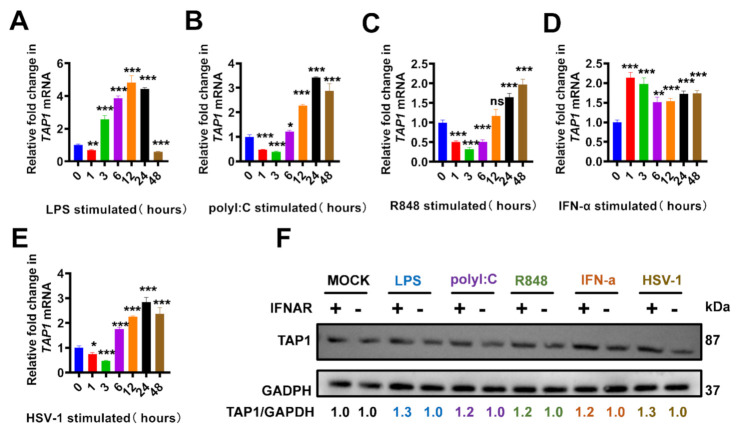
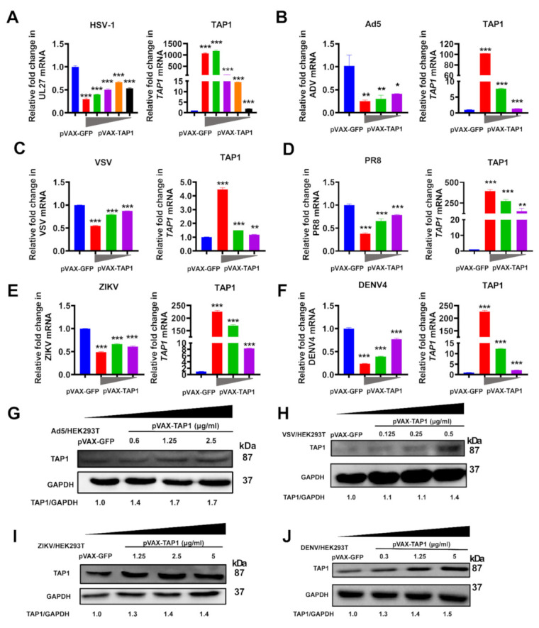
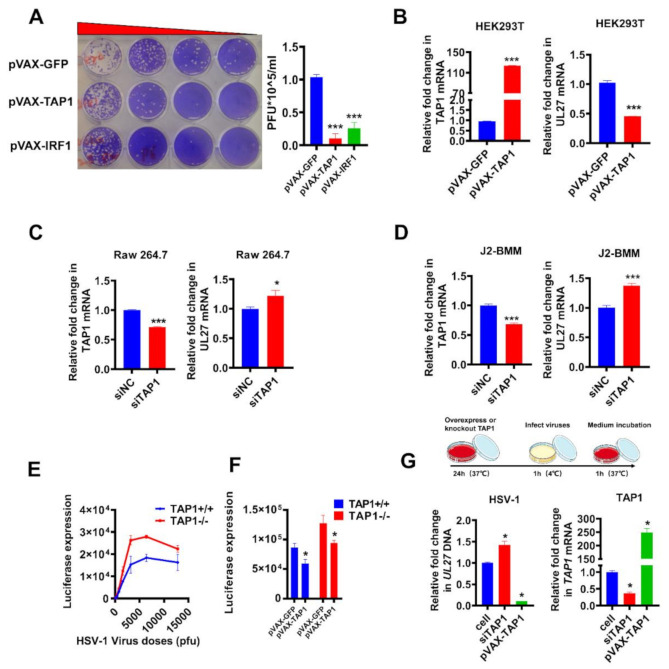
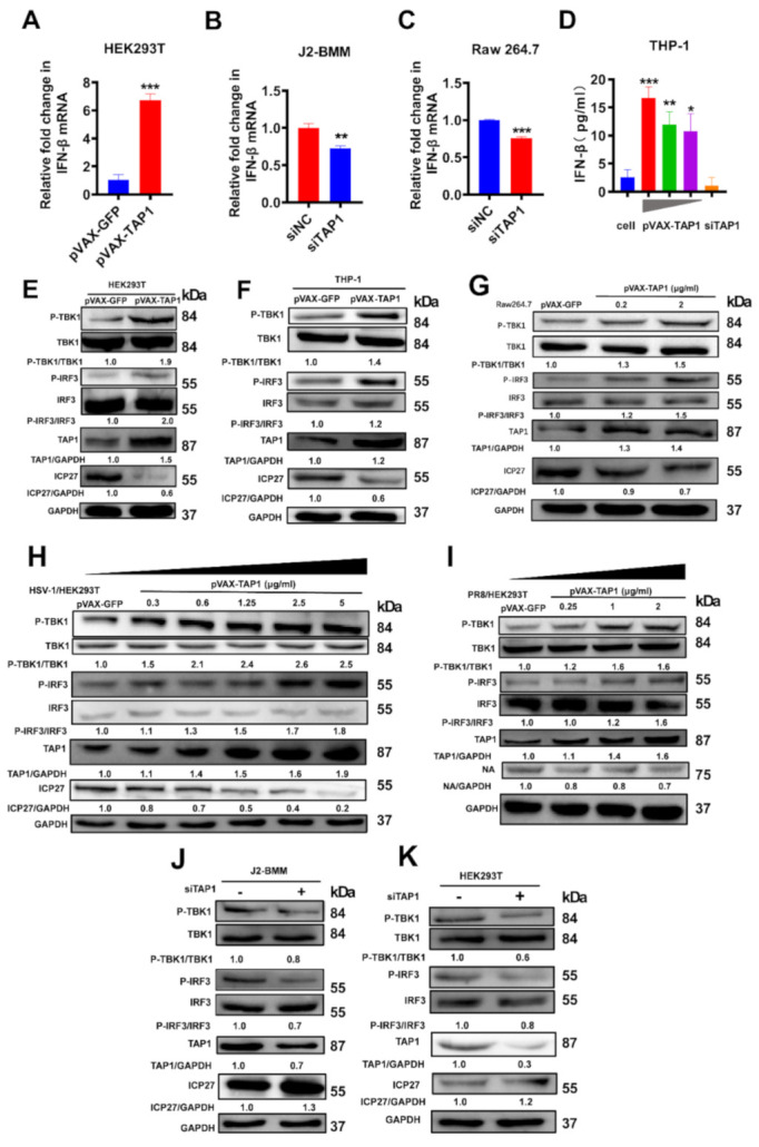
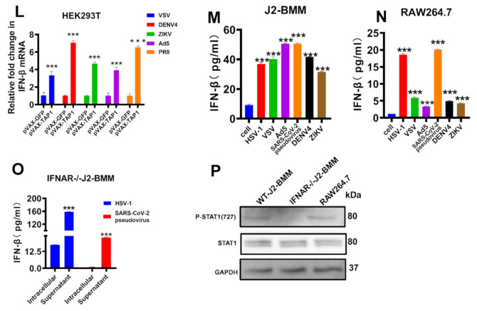
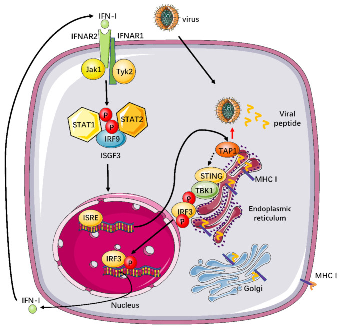
Deeply understanding the virus-host interaction is a prerequisite for developing effective anti-viral strategies. Traditionally, the transporter associated with antigen processing type 1 (TAP1) is critical for antigen presentation to regulate adaptive immunity. However, its role in controlling viral infections through modulating innate immune signaling is not yet fully understood. In the present study, we reported that TAP1, as a product of interferon-stimulated genes (ISGs), had broadly antiviral activity against various viruses such as herpes simplex virus 1 (HSV-1), adenoviruses (AdV), vesicular stomatitis virus (VSV), dengue virus (DENV), Zika virus (ZIKV), and influenza virus (PR8) etc. This antiviral activity by TAP1 was further confirmed by series of loss-of-function and gain-of-function experiments. Our further investigation revealed that TAP1 significantly promoted the interferon (IFN)-β production through activating the TANK binding kinase-1 (TBK1) and the interferon regulatory factor 3 (IRF3) signaling transduction. Our work highlighted the broadly anti-viral function of TAP1 by modulating innate immunity, which is independent of its well-known function of antigen presentation. This study will provide insights into developing novel vaccination and immunotherapy strategies against emerging infectious diseases.

Keywords: broadly antiviral activity; innate immunity; interferon-stimulated genes (ISGs); transporter associated with antigen processing 1 (TAP1); type I interferons (IFN-Is).


Conflict of interest statement

The authors declare no conflict of interest.

Figures

Figure 1
Figure 1
The upregulated TAP1 in response to Toll-like receptors (TLRs) agonists and viral infections. (AE) Raw 264.7 cells were stimulated with LPS (1 μg/mL), polyI:C (25 μg/mL), R848 (100 nM), IFN-α (2000 U/mL), and HSV-1 (MOI = 0.25), respectively, and then the expression of TAP1 gene was measured by RT-qPCR at different timepoints. The expression level of mRNA was normalized to the expression of β-actin, and the data from at least triplicates were shown as the mean ± SD. * p < 0.05, ** p < 0.01, *** p < 0.001. (F) Wild type bone marrow-derived macrophage (WT-J2-BMM) cells or interferon α receptor-deficient (IFNAR−/−)-J2-BMM cells were stimulated with LPS (1 μg/mL), polyI:C (25 μg/mL), R848 (100 nM), R848 (100 nM), IFN-α (2000 U/mL), and HSV-1(MOI = 0.25) for 24 h, respectively, and then the expression of TAP1 protein was measured by Western blot analysis. GAPDH was used as intern control. The relative ratios of TAP1 and GAPDH were marked at the bottom of the pictures.
Figure 2
Figure 2
Broadly antiviral activities of TAP1. (A) HEK293T cells were transfected with TAP1-expressing plasmid in different concentrations (2.5 μg/mL, 1.25 μg/mL, 0.6 μg/mL, and 0.3 μg/mL) for 24 h, followed by HSV-1 infection (MOI = 1) for 8 h. The expression level of TAP1 and HSV-1 UL27 was quantified by RT-qPCR. (B) HEK293T cells were transfected with TAP1-expressing plasmid in different concentrations (2.5 μg/mL, 1.25 μg/mL, and 0.6 μg/mL) for 24 h, followed by Ad5 infection (MOI = 1) for 8 h. The expression level of TAP1 and Ad5 was quantified by RT-qPCR. (C) HEK293T cells were transfected with TAP1-expressing plasmid in different concentrations (0.5 μg/mL, 0.25 μg/mL, and 0.125 μg/mL) for 24 h, followed by VSV infection (MOI = 1) for 8 h. The expression level of TAP1 and VSV was quantified by RT-qPCR. (D) HEK293T cells were transfected with TAP1-expressing plasmid in different concentrations (2 μg/mL, 1 μg/mL, and 0.25 μg/mL) for 24 h, followed by PR8 infection (MOI = 1) for 8 h. The expression level of TAP1 and PR8 was quantified by RT-qPCR (E) HEK293T cells were transfected with TAP1-expressing plasmid in different concentrations (5 μg/mL, 2.5 μg/mL, and 1.25 μg/mL) for 24 h, followed by ZIKV infection (MOI = 1) for 8 h. The expression level of TAP1 and ZIKV was quantified by RT-qPCR. (F) HEK293T cells were transfected with TAP1-expressing plasmid in different concentrations (5 μg/mL, 1.25 μg/mL, and 0.3 μg/mL) for 24 h, followed by DENV4 infection (MOI = 1) for 8 h. The expression level of TAP1 and DENV4 was quantified by RT-qPCR. The expression level of mRNA was normalized to the expression of β-actin, and data from at least triplicates were shown as the mean ± SD. * p < 0.05, ** p < 0.01, and *** p < 0.001. (GJ) HEK293T cells were transfected with TAP1-expressing plasmid in indicated concentrations for 24 h, followed by Ad5, VSV, ZIKV, and DENV4 infection (MOI = 1) for 8 h, respectively. The expression level of TAP1 and GAPDH was quantified by Western blot, and the relative ratios of TAP1 and GAPDH were labeled at the bottom of the pictures. GAPDH was used as the internal control.
Figure 3
Figure 3
Validation of viral inhibition by loss-of-function and gain-of-function experiments. (A)Vero cells were transfected with plasmids (1 μg/mL) expressing GFP, TAP1, and IRF1 for 24 h, respectively, followed by HSV-1 infection (MOI = 0.25) for 8 h. Then, the titer of HSV-1 in the supernatant was quantified by plaque assay. (B) HEK293T cells were transfected with plasmids (1 μg/mL) expressing GFP and TAP1 for 24 h, respectively, followed by HSV-1 infection (MOI = 0.25) for 8 h. Then, the expression of TAP1 and HSV-1 UL27 were quantified by RT-qPCR. (C,D) Raw 264.7 and WT-J2-BMM cells were transfected with small interfering RNA (siRNA) (siNC as negative control) for 24 h, followed by HSV-1 infection (MOI = 0.25) for 8 h. Then, the expression of TAP1 and HSV-1 UL27 were quantified by RT-qPCR. The expression level of mRNA was normalized to the expression of β-actin. (E) TAP1-wildtype (TAP1+/+) or TAP1-knockout (TAP1−/−) fibroblast cells were infected with HSV-1, and subsequently, the Renilla luciferase activity in cell lysates was determined at 8 h post infection. (F) TAP1+/+ or T TAP1−/− fibroblast cells were transfected with or without TAP1-expressing plasmid (1 μg/mL) for 24 h, followed by HSV-1 infection (MOI = 0.25) for 8 h, and then the Renilla luciferase activity in cell lysates was determined. (G) HEK293 cells were transfected with plasmids (1 μg/mL) expressing TAP1 or siTAP1 for 24 h, respectively, followed by HSV-1 incubation (MOI = 1) for 1 h at 4 °C. Then, aspirated and discarded the supernatant, washed with PBS three times, added culture medium, incubated at 37 °C for 1 h, and the expression of HSV-1 (UL27) and TAP1 were quantified by qPCR. The expression level was normalized to the expression of β-actin, and the data from at least triplicates were shown as the mean ± SD. * p < 0.05, and *** p < 0.001.
Figure 4
Figure 4
TAP1 significantly promoted the interferon (IFN)-β production through activating the TANK binding kinase-1 (TBK1) and the interferon regulatory factor 3 (IRF3) signaling transduction. (A) HEK293T cells were transfected with plasmids expressing GFP or TAP1 (1 μg/mL) for 24 h, respectively, followed by HSV-1 infection (MOI = 0.25) for 8 h. Then, the expression of IFN-β was quantified by RT-qPCR. (B) WT-J2-BMM cells were transfected with siNC, siTAP1 for 24 h, followed by HSV-1 infection (MOI = 0.25) for 8 h. Then, the expression of IFN-β was quantified by RT-qPCR. (C) Raw264.7 cells were transfected with siNC, siTAP1 for 24 h, followed by HSV-1 infection (MOI = 0.25) for 8 h. Then, the expression of IFN-β was quantified by RT-qPCR. (D) THP-1 cells were transfected with TAP1-expressing plasmid in different concentration (5 μg/mL, 1.25 μg/mL, 0.3 μg/mL) for 24 h, followed by HSV-1 infection (MOI = 0.25). Then, the expression of IFN-β in cell lysates was determined by ELISA kits. (EG) HEK293T, THP-1 or Raw 264.7 cells were transfected with plasmids expressing GFP or TAP1 (1 μg/mL) for 24 h respectively, followed by HSV-1 infection (MOI = 0.25) for 8 h. Then, the expression of targeted proteins was detected by Western blot. The relative ratios of targeted protein and GAPDH were marked at the bottom of the pictures. (H,I) HEK293T cells were transfected with plasmids expressing GFP or TAP1 for 24 h, followed by viral infections (HSV-1 or PR8) for 24 h at MOI of 1. Then, the expression of targeted proteins was detected by Western blot. The relative ratios of targeted protein and GAPDH were marked at the bottom of the pictures. (J,K) WT-J2-BMM or HEK293T cells were transfected with siNC or siTAP1 for 24 h, followed by HSV-1 infection (MOI = 0.25) for 8 h. Then, the expression of targeted proteins was detected by Western blot. The relative ratios of targeted protein and GAPDH were marked at the bottom of the pictures. (L) HEK293T cells were transfected with or without TAP1-expressing plasmid for 24 h, followed by viral infections for 8 h at MOI of 1, including VSV, ZIKV, DENV4, Ad5 and PR8. Then, the expression of IFN-β was quantified by RT-qPCR. (M,N) WT-J2-BMM or Raw 264.7 cells were transfected with TAP1-expressing plasmid for 24 h, followed by viral infections for 24 h at 1 MOI, including HSV-1, VSV, Ad5, SARS-CoV-2 pseudovirus, ZIKV, DENV4. Then, the expression of IFN-β in cell lysates was determined by ELISA kits. (O) (IFNAR−/−)-J2-BMM cells were transfected with TAP1-expressing plasmid for 24 h, followed by HSV-1 or SARS-CoV-2 pseudovirus infection for 24 h. Then, the expression of IFN-β in cell lysates and supernatant was determined by ELISA kits. (P) WT-J2-BMM, (IFNAR−/−)-J2-BMM, and Raw 264.7 cells were transfected with TAP1-expressing plasmid for 24 h, followed by HSV-1 infection for 24 h. Then, the level of STAT1 phosphorylation and total STAT1 was measured by Western blot. The data from at least triplicates were shown as the mean ± SD. * p < 0.05, ** p < 0.01 and *** p < 0.001.
Figure 4
Figure 4
TAP1 significantly promoted the interferon (IFN)-β production through activating the TANK binding kinase-1 (TBK1) and the interferon regulatory factor 3 (IRF3) signaling transduction. (A) HEK293T cells were transfected with plasmids expressing GFP or TAP1 (1 μg/mL) for 24 h, respectively, followed by HSV-1 infection (MOI = 0.25) for 8 h. Then, the expression of IFN-β was quantified by RT-qPCR. (B) WT-J2-BMM cells were transfected with siNC, siTAP1 for 24 h, followed by HSV-1 infection (MOI = 0.25) for 8 h. Then, the expression of IFN-β was quantified by RT-qPCR. (C) Raw264.7 cells were transfected with siNC, siTAP1 for 24 h, followed by HSV-1 infection (MOI = 0.25) for 8 h. Then, the expression of IFN-β was quantified by RT-qPCR. (D) THP-1 cells were transfected with TAP1-expressing plasmid in different concentration (5 μg/mL, 1.25 μg/mL, 0.3 μg/mL) for 24 h, followed by HSV-1 infection (MOI = 0.25). Then, the expression of IFN-β in cell lysates was determined by ELISA kits. (EG) HEK293T, THP-1 or Raw 264.7 cells were transfected with plasmids expressing GFP or TAP1 (1 μg/mL) for 24 h respectively, followed by HSV-1 infection (MOI = 0.25) for 8 h. Then, the expression of targeted proteins was detected by Western blot. The relative ratios of targeted protein and GAPDH were marked at the bottom of the pictures. (H,I) HEK293T cells were transfected with plasmids expressing GFP or TAP1 for 24 h, followed by viral infections (HSV-1 or PR8) for 24 h at MOI of 1. Then, the expression of targeted proteins was detected by Western blot. The relative ratios of targeted protein and GAPDH were marked at the bottom of the pictures. (J,K) WT-J2-BMM or HEK293T cells were transfected with siNC or siTAP1 for 24 h, followed by HSV-1 infection (MOI = 0.25) for 8 h. Then, the expression of targeted proteins was detected by Western blot. The relative ratios of targeted protein and GAPDH were marked at the bottom of the pictures. (L) HEK293T cells were transfected with or without TAP1-expressing plasmid for 24 h, followed by viral infections for 8 h at MOI of 1, including VSV, ZIKV, DENV4, Ad5 and PR8. Then, the expression of IFN-β was quantified by RT-qPCR. (M,N) WT-J2-BMM or Raw 264.7 cells were transfected with TAP1-expressing plasmid for 24 h, followed by viral infections for 24 h at 1 MOI, including HSV-1, VSV, Ad5, SARS-CoV-2 pseudovirus, ZIKV, DENV4. Then, the expression of IFN-β in cell lysates was determined by ELISA kits. (O) (IFNAR−/−)-J2-BMM cells were transfected with TAP1-expressing plasmid for 24 h, followed by HSV-1 or SARS-CoV-2 pseudovirus infection for 24 h. Then, the expression of IFN-β in cell lysates and supernatant was determined by ELISA kits. (P) WT-J2-BMM, (IFNAR−/−)-J2-BMM, and Raw 264.7 cells were transfected with TAP1-expressing plasmid for 24 h, followed by HSV-1 infection for 24 h. Then, the level of STAT1 phosphorylation and total STAT1 was measured by Western blot. The data from at least triplicates were shown as the mean ± SD. * p < 0.05, ** p < 0.01 and *** p < 0.001.
Figure 5
Figure 5
Working model to illustrate the TAP1-involved innate signal pathway against broadly viral infections. TAP1 plays a critical factor in the profound virus-host interactions, especially in innate and adaptive immunity. TAP1 can regulate adaptive immunity through promoting the major histocompatibility complex (MHC) class I-mediated antigen presentation. Additionally, our study also demonstrated that TAP1 could promote type I interferon production through activating the TBK1-IRF3 signal pathway. Subsequently, Type I IFNs can bind to IFNAR2, then recruit IFNAR1, and consequently, form a signaling-competent ternary complex to activate the transcription factor IRF9 and ISGF3 complex, which is comprised of phosphorylated STAT1 and STAT2, through the JAK-STAT signal pathway. The activated ISGF3 can translocate to the nucleus and bind to IFN-stimulated response elements (ISREs) in the upstream promoter regions of ISGs, which can encode the numerous proteins with potent antiviral activities.

Similar articles

Cited by

References

    1. Ma Y., Su X.Z., Lu F. The Roles of Type I Interferon in Co-infections With Parasites and Viruses, Bacteria, or Other Parasites. Front. Immunol. 2020;11:1805. doi: 10.3389/fimmu.2020.01805. - DOI - PMC - PubMed
    1. Kienes I., Weidl T., Mirza N., Chamaillard M., Kufer T.A. Role of NLRs in the Regulation of Type I Interferon Signaling, Host Defense and Tolerance to Inflammation. Int. J. Mol. Sci. 2021;22:1301. doi: 10.3390/ijms22031301. - DOI - PMC - PubMed
    1. Schoggins J.W. Interferon-Stimulated Genes: What Do They All Do? Annu. Rev. Virol. 2019;6:567–584. doi: 10.1146/annurev-virology-092818-015756. - DOI - PubMed
    1. Abele R., Tampe R. The ABCs of immunology: Structure and function of TAP, the transporter associated with antigen processing. Physiology. 2004;19:216–224. doi: 10.1152/physiol.00002.2004. - DOI - PubMed
    1. Parcej D., Tampé R. ABC proteins in antigen translocation and viral inhibition. Nat. Chem. Biol. 2010;6:572–580. doi: 10.1038/nchembio.410. - DOI - PubMed

MeSH terms

LinkOut - more resources