

The Contribution of Microglia to Neuroinflammation in Parkinson's Disease
- PMID: 33925154
- PMCID: PMC8125756
- DOI: 10.3390/ijms22094676
The Contribution of Microglia to Neuroinflammation in Parkinson's Disease
Abstract
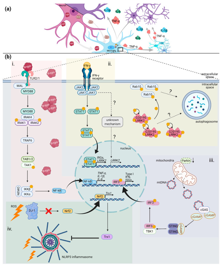
With the world's population ageing, the incidence of Parkinson's disease (PD) is on the rise. In recent years, inflammatory processes have emerged as prominent contributors to the pathology of PD. There is great evidence that microglia have a significant neuroprotective role, and that impaired and over activated microglial phenotypes are present in brains of PD patients. Thereby, PD progression is potentially driven by a vicious cycle between dying neurons and microglia through the instigation of oxidative stress, mitophagy and autophagy dysfunctions, a-synuclein accumulation, and pro-inflammatory cytokine release. Hence, investigating the involvement of microglia is of great importance for future research and treatment of PD. The purpose of this review is to highlight recent findings concerning the microglia-neuronal interplay in PD with a focus on human postmortem immunohistochemistry and single-cell studies, their relation to animal and iPSC-derived models, newly emerging technologies, and the resulting potential of new anti-inflammatory therapies for PD.
Keywords: Parkinson’s disease; animal models; brain; iPSC; microglia; neurodegeneration; neuroinflammation.
Conflict of interest statement
The authors declare no conflict of interest.
Figures

References
Publication types
MeSH terms
Substances
Grants and funding
LinkOut - more resources
Full Text Sources
Other Literature Sources
Medical
