. 2021 Apr 28;11(5):1157.
doi: 10.3390/nano11051157.

Collagen Film Activation with Nanoscale IKVAV-Capped Dendrimers for Selective Neural Cell Response

Affiliations

Collagen Film Activation with Nanoscale IKVAV-Capped Dendrimers for Selective Neural Cell Response

Jessica J Kim et al. Nanomaterials (Basel). .

Abstract

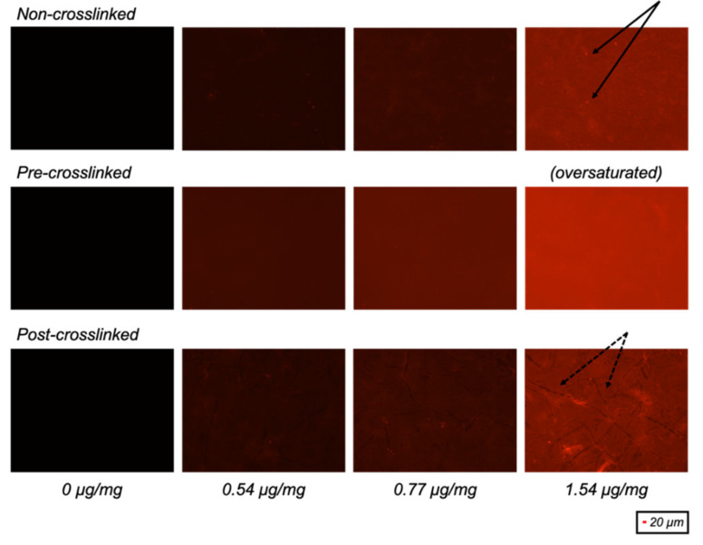
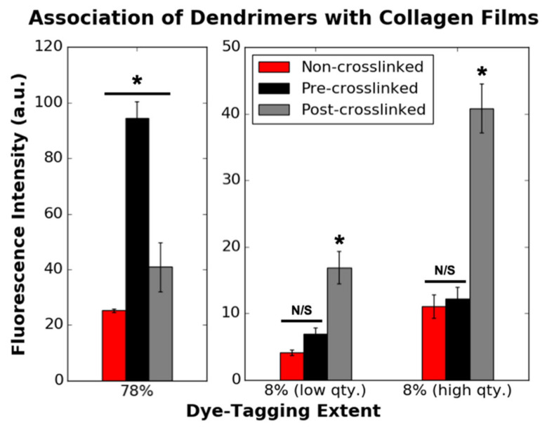
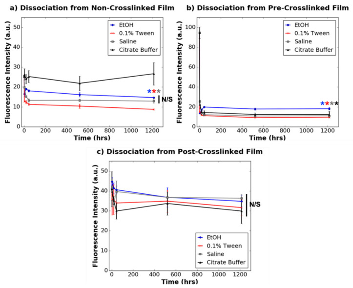
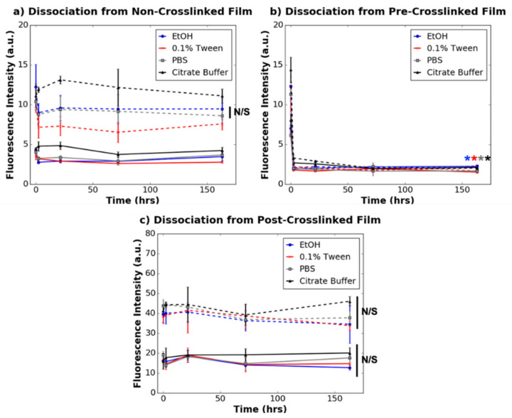
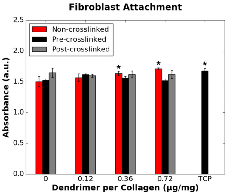
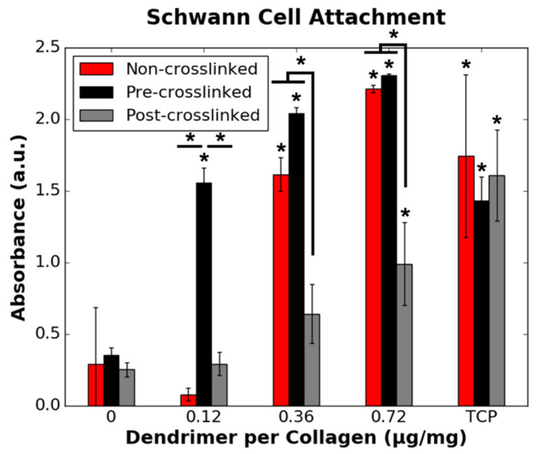
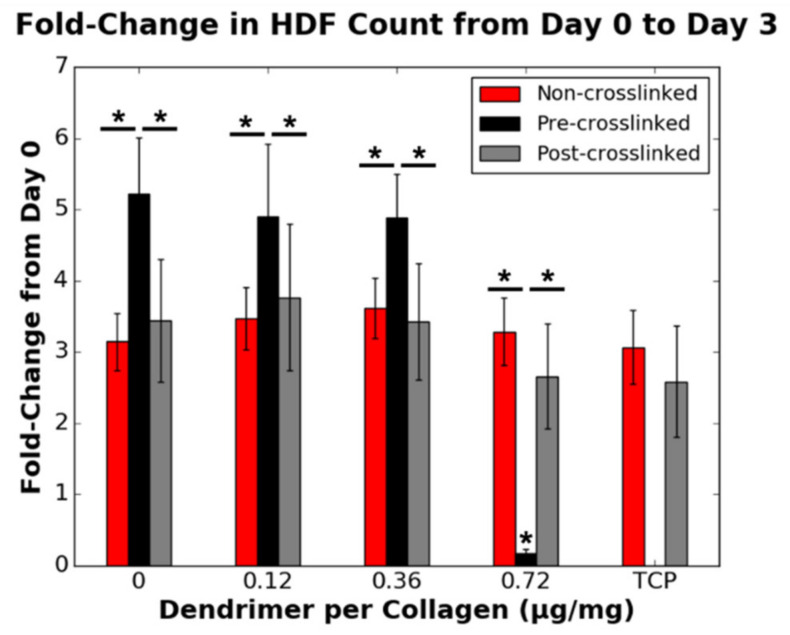
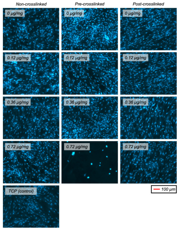
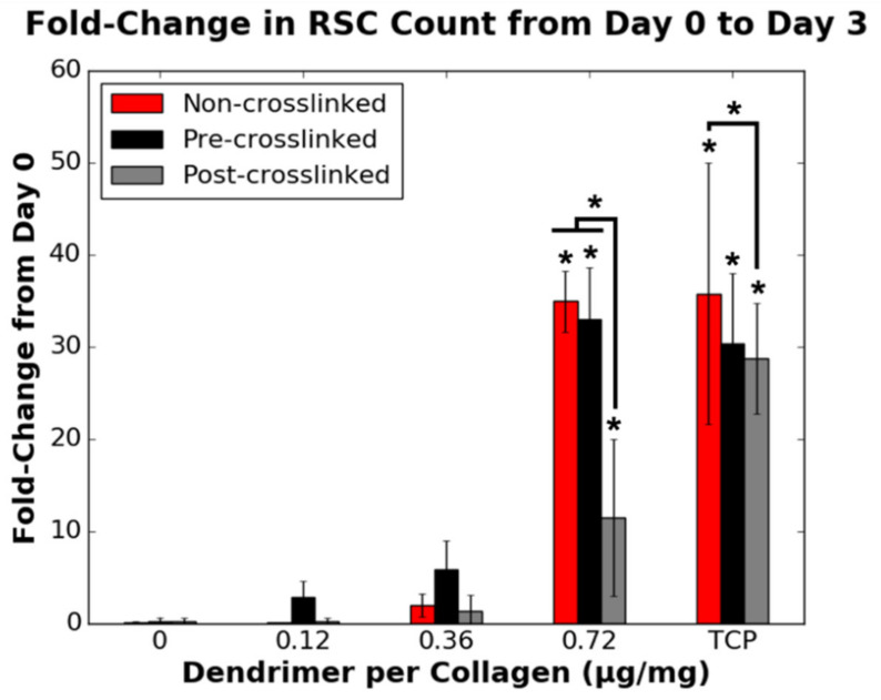
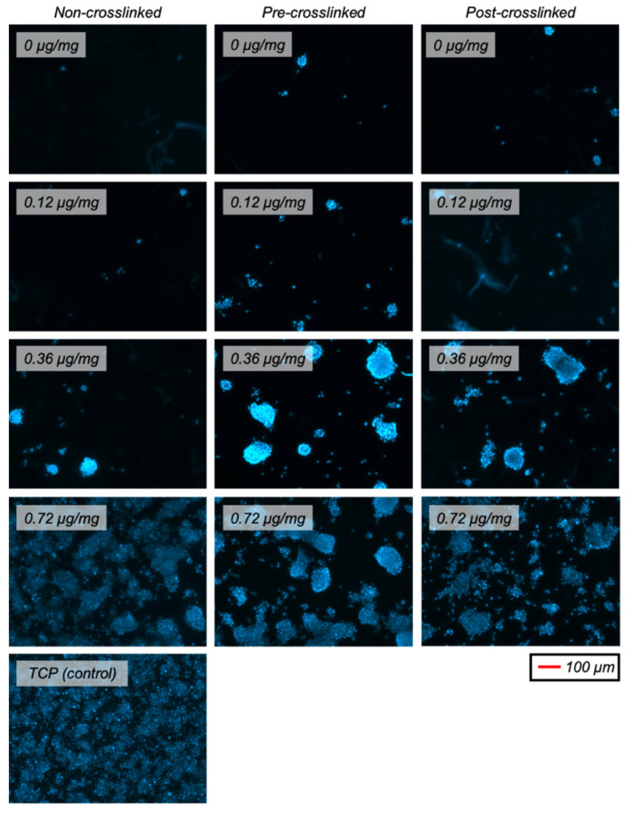
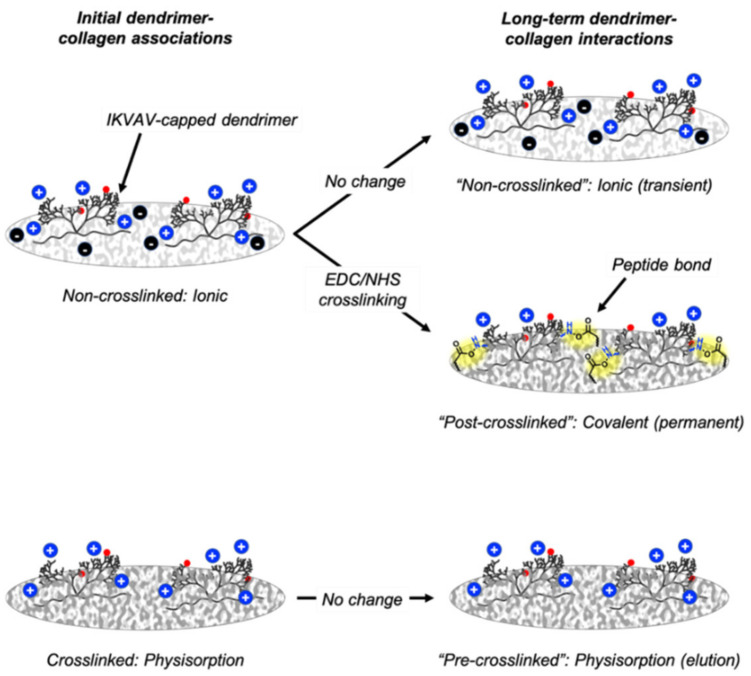
Biocompatible neural guidance conduits are alternatives to less abundant autologous tissue grafts for small nerve gap injuries. To address larger peripheral nerve injuries, it is necessary to design cell selective biomaterials that attract neuronal and/or glial cells to an injury site while preventing the intrusion of fibroblasts that cause inhibitory scarring. Here, we investigate a potential method for obtaining this selective cellular response by analysing the responses of rat Schwann cells and human dermal fibroblasts to isoleucine-lysine-valine-alanine-valine (IKVAV)-capped dendrimer-activated collagen films. A high quantity of nanoscale IKVAV-capped dendrimers incorporated onto pre-crosslinked collagen films promoted rat Schwann cell attachment and proliferation, and inhibited human dermal fibroblast proliferation. In addition, while pre-crosslinked dendrimer-activated films inhibited fibroblast proliferation, non-crosslinked dendrimer-activated films and films that were crosslinked after dendrimer-activation (post-crosslinked films) did not. The different cellular responses to pre-crosslinked and post-crosslinked films highlight the importance of having fully exposed, non-covalently bound biochemical motifs (pre-crosslinked films) directing certain cellular responses. These results also suggest that high concentrations of nanoscale IKVAV motifs can inhibit fibroblast attachment to biological substrates, such as collagen, which inherently attract fibroblasts. Therefore, this work points toward the potential of IKVAV-capped dendrimer-activated collagen biomaterials in limiting neuropathy caused by fibrotic scarring at peripheral nerve injury sites.

Keywords: IKVAV pentapeptide; bioactivated materials; collagen; dendrimers.


Conflict of interest statement

The authors declare no conflict of interest.

Figures

Figure 1
Figure 1
Fluorescence images of non-crosslinked, pre-crosslinked and post-crosslinked collagen films with different quantities of 78% dye-conjugated dendrimer activation (µg dendrimer per mg collagen). Presence of overexposed bright particulates (solid) and underexposed collagen “coils” (dashed) are indicated by black arrows.
Figure 2
Figure 2
Initial association of 78% (0.77 µg/mg loading concentration) (n = 4) and 8% (n = 12) dye-tagged dendrimers with non-crosslinked, pre-crosslinked and post-crosslinked films. Eight percent (low qty.) and 8% (high qty.) correspond to 0.74 and 1.49 µg/mg loading concentrations (µg dendrimer per mg collagen), respectively. This data corresponds to the 0 h values in Figure 3 and Figure 4. N/S denotes p ≥ 0.05. * indicates statistically significant difference (p ≤ 0.001) from the two other substrate varieties with dendrimers with equivalent dye-tagging extents.
Figure 3
Figure 3
Dissociation of 78% dye-tagged dendrimers (0.77 µg/mg loading concentration) from (a) non-crosslinked films, (b) pre-crosslinked films, and (c) post-crosslinked films (n = 3 for all time points except for time point 0, which was n = 4). N/S denotes p ≥ 0.05. * denotes statistically significant difference (p ≤ 0.01) between the data point annotated (colour-coded) and the respective 0 h value for each collagen substrate. Statistical significance is only denoted for the final time point for clarity.
Figure 4
Figure 4
Dissociation of 8% dye-tagged dendrimers from (a) non-crosslinked films, (b) pre-crosslinked films, and (c) post-crosslinked films. Solid and dotted lines correspond to 0.74 µg/mg and 1.49 µg/mg dendrimer loading concentrations, respectively. N/S denotes p ≥ 0.05. * denotes statistically significant difference (p ≤ 0.01) between the data point annotated (colour-coded) and the respective 0 h value for each collagen substrate. Statistical significance is only depicted for 1.49 µg/mg dendrimer quantities for non-crosslinked and pre-crosslinked films and only for the final time point for clarity.
Figure 5
Figure 5
Effect of the quantity of IKVAV-capped dendrimers added to the various collagen films on HDF binding. HDFs were seeded onto non-collagen coated wells (TCP controls) on the same 48-well plate as pre-crosslinked films. * denotes statistically significant difference (p ≤ 0.05) between the data point annotated and the respective 0 µg/mg value for each collagen substrate.
Figure 6
Figure 6
Effect of the quantity of IKVAV-capped dendrimers added to the various collagen films on RSC binding. RSCs were seeded onto non-collagen coated wells (TCP controls) on the same 48-well plates as each collagen substrate variety. * denotes statistically significant difference (p ≤ 0.05) between the data point annotated and the respective 0 µg/mg value for each collagen substrate, unless indicated otherwise.
Figure 7
Figure 7
Effect of the quantity of IKVAV-capped dendrimers added to the various collagen substrates on the fold-change in HDF cell count from day 0 to day 3 (n = 9). A fold-change of 1 is equivalent to no change in cell density. HDFs were seeded onto non-collagen coated wells (TCP controls) on the same 48-well plates as non-crosslinked and post-crosslinked films. * denotes statistically significant difference (p ≤ 0.01) between the data point annotated and the respective 0 µg/mg value for each collagen substrate, unless indicated otherwise.
Figure 8
Figure 8
Representative fluorescent images (DAPI-stained nuclei) of HDFs on various collagen (or TCP) substrates after 3 days in culture.
Figure 9
Figure 9
Effect of the quantity of IKVAV-capped dendrimers added to the various collagen substrates on the fold-change in RSC cell count from day 0 to day 3 (n = 9). A fold-change of 1 is equivalent to no change in cell density. RSCs were seeded onto non-collagen coated wells (TCP controls) on the same 48-well plates as each collagen substrate variety. * denotes statistically significant difference (p ≤ 0.001) between the data point annotated and the respective 0 µg/mg value for each collagen substrate, unless indicated otherwise.
Figure 10
Figure 10
Representative fluorescent images (DAPI-stained nuclei) of RSCs on various collagen (or TCP) substrates after 3 days in culture.
Figure 11
Figure 11
Chemical interactions between dendrimers and non-crosslinked, pre-crosslinked and post-crosslinked films. Red spheres depict Atto dye, blue spheres depict positively charged free amines on IKVAV-capped dendrimers, black spheres depict negatively charged glutamate/aspartate residues on non-crosslinked collagen, and yellow highlights dendrimer-collagen covalent bonds.

References

    1. Daly W., Yao L., Zeugolis D., Windebank A., Pandit A. A biomaterials approach to peripheral nerve regeneration: Bridging the peripheral nerve gap and enhancing functional recovery. J. R. Soc. Interface. 2012;9:202–221. doi: 10.1098/rsif.2011.0438. - DOI - PMC - PubMed
    1. Kubiak C.A., Kung T.A., Brown D.L., Cederna P.S., Kemp S.W.P. State-of-the-Art techniques in treating peripheral nerve injury. Plast. Reconstr. Surg. 2018;141:702–710. doi: 10.1097/PRS.0000000000004121. - DOI - PubMed
    1. Li H., Wong C., Li W., Ruven C., He L., Wu X., Lang B.T., Silver J., Wu W. Enhanced regeneration and functional recovery after spinal root avulsion by manipulation of the proteoglycan receptor PTPσ. Sci. Rep. 2015;5:1–14. doi: 10.1038/srep14923. - DOI - PMC - PubMed
    1. Wang M.L., Rivlin M., Graham J.G., Beredjiklian P.K. Peripheral nerve injury, scarring, and recovery. Connect. Tissue Res. 2019;60:3–9. doi: 10.1080/03008207.2018.1489381. - DOI - PubMed
    1. Dreesmann L., Mittnacht U., Lietz M., Schlosshauer B. Nerve fibroblast impact on Schwann cell behavior. Eur. J. Cell Biol. 2009;88:285–300. doi: 10.1016/j.ejcb.2009.01.001. - DOI - PubMed

LinkOut - more resources