Review
. 2021 Apr 27;22(9):4591.
doi: 10.3390/ijms22094591.

On Iron Metabolism and Its Regulation

Affiliations
Review

On Iron Metabolism and Its Regulation

Anne-Cathrine S Vogt et al. Int J Mol Sci. .

Abstract

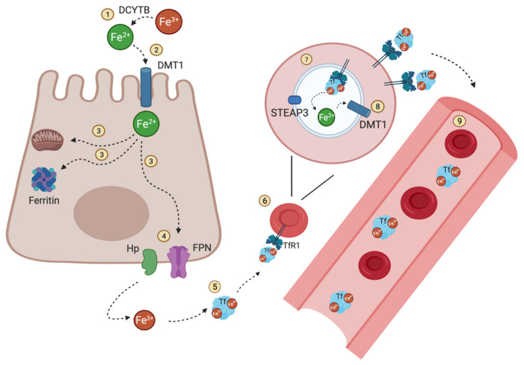
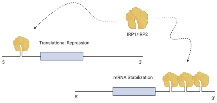
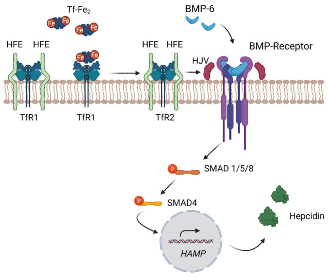
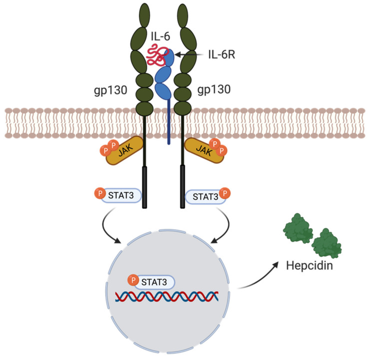
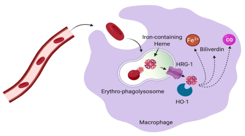
Iron is a critical metal for several vital biological processes. Most of the body's iron is bound to hemoglobin in erythrocytes. Iron from senescent red blood cells is recycled by macrophages in the spleen, liver and bone marrow. Dietary iron is taken up by the divalent metal transporter 1 (DMT1) in enterocytes and transported to portal blood via ferroportin (FPN), where it is bound to transferrin and taken up by hepatocytes, macrophages and bone marrow cells via transferrin receptor 1 (TfR1). While most of the physiologically active iron is bound hemoglobin, the major storage of most iron occurs in the liver in a ferritin-bound fashion. In response to an increased iron load, hepatocytes secrete the peptide hormone hepcidin, which binds to and induces internalization and degradation of the iron transporter FPN, thus controlling the amount of iron released from the cells into the blood. This review summarizes the key mechanisms and players involved in cellular and systemic iron regulation.

Keywords: hepcidin; iron; macrophages.


Conflict of interest statement

The authors declare no conflict of interest.

Figures

Figure 1
Figure 1
Iron distribution and circulation. Nonheme dietary iron Fe3+ is reduced to Fe2+ by the iron reducing DCYTB (1) prior to its uptake at the apical membrane of enterocytes via DMT1(2). Fe2+ can then be directly used for intracellular mechanisms, stored when bound to ferritin or released directly into the circulation (3). (4) Therefore, reduced iron Fe2+ is transported by ferroportin (FPN), the only known iron exporter so far, and then oxidized by hephaestin Hp to be then bound to Tf (5). Most of the iron present in the circulation is bound to Tf. As a result, erythrocyte precursors (erythroblasts) take up this transferrin-bound iron via TfR1(6). Fe3+ bound to transferrin is reduced in the endosome by ferrireductase STEAP3 to Fe2+ (7) where it is exported via DMT1 (8) into the cytosol and enters the labile iron pool. Mature RBCs circulate in the blood for around 120 days (9) until they are removed from the circulation during erythrophagocytosis. The illustration was created using BioRender.com, (accessed on 3 April 2021).
Figure 2
Figure 2
Cellular iron regulation. The intracellular iron pool is regulated by the binding of IRP1 and IRP2 to IRE. IRP1 and IRP2 are RNA binding proteins that interact with IRE to control the translation of proteins involved in iron metabolism. IRPs are either present at the 3′ UTR or the 5′ UTR of the target mRNA. When an IRP binds to a single IRE at the 5′ UTR, mRNA translation is repressed. On the other hand, the binding of IRP to IRE at the 3′ UTR stabilizes the transcript and leads to increased mRNA translation. The illustration was created using BioRender.com, (accessed on 3 April 2021).
Figure 3
Figure 3
Regulation of Hepcidin Expression. Circulating hepcidin regulates the amount of iron released into the blood from macrophages and especially hepatocytes. Decreased hepcidin expression occurs when the rate of erythropoiesis increases (e.g., in response to anemia), leading to increased ferroportin expression causing increased iron transfer into blood. In contrast, hepcidin expression is increased by elevated plasma iron (Tf-Fe2) or inflammation to counteract an oversaturation of Tf or iron loss, preventing the formation of cytotoxic NTBI. The liver directly senses circulating iron bound to Tf or indirectly in response to iron-induced BMP6. Increased hepatic iron levels induce the expression of BMP6. BMP6 stimulates hepcidin expression by binding to the BMP receptor and HJV, leading to intracellular signaling via SMAD proteins, coupled with SMAD4 translocating to the nucleus and inducing hepcidin expression. The liver directly senses circulating iron by expression of TfR1 and TfR2. With increasing serum iron levels, Tf-Fe2 binds to TfR1 and HFE binding to TfR2 is induced. This complex interacts with HJV and enhances the BMP signaling pathway, leading to hepcidin transcription. The illustration was created using BioRender.com, (accessed on 22 April 2021).
Figure 4
Figure 4
Inflammation triggering hepcidin expression. The inflammatory cytokine interleukin 6 (IL-6) can lead to hepcidin induction via the IL-6R/STAT3 pathway. The binding of IL-6 to its corresponding receptor IL-6R leads to the downstream phosphorylation of STAT3 via JAK1/2. After its phosphorylation, STAT3 will translocate into the nucleus binding to the hepcidin promotor inducing hepcidin expression. The illustration was created using BioRender.com, (accessed on 3 April 2021).
Figure 5
Figure 5
Erythrophagocytosis by macrophages. Macrophages in the red pulp of the spleen destroy senescent red blood cells and recycle the stored iron to be further incorporated into maturating RBCs during erythropoiesis. Senescent RBCs are engulfed into the phagolysosome of macrophages. Within the erythro-phagolysosome, RBCs are digested resulting in the breakdown of hemoglobin. Iron-containing heme is transported into the cytosol via HRG1. In the cytosol, iron bound to heme is processed by HO-1, resulting in the release of Fe2+, biliverdin and CO. The illustration was created using BioRender.com, (accessed on 3 April 2021).

References

    1. Sukhbaatar N., Weichhart T. Iron Regulation: Macrophages in Control. Pharmaceuticals. 2018;11:137. doi: 10.3390/ph11040137. - DOI - PMC - PubMed
    1. Dev S., Babitt J.L. Overview of Iron Metabolism in Health and Disease. Hemodial. Int. 2017;21:S6–S20. doi: 10.1111/hdi.12542. - DOI - PMC - PubMed
    1. Silva B., Faustino P. An Overview of Molecular Basis of Iron Metabolism Regulation and the Associated Pathologies. Biochim. Biophys. Acta Mol. Basis Dis. 2015;1852:1347–1359. doi: 10.1016/j.bbadis.2015.03.011. - DOI - PubMed
    1. Schwartz A.J., Converso-Baran K., Michele D.E., Shah Y.M. A Genetic Mouse Model of Severe Iron Deficiency Anemia Reveals Tissue-Specific Transcriptional Stress Responses and Cardiac Remodeling. J. Biol. Chem. 2019;294:14991–15002. doi: 10.1074/jbc.RA119.009578. - DOI - PMC - PubMed
    1. Jomova K., Valko M. Advances in Metal-Induced Oxidative Stress and Human Disease. Toxicology. 2011;283:65–87. doi: 10.1016/j.tox.2011.03.001. - DOI - PubMed