. 2021 Apr 26;9(5):474.
doi: 10.3390/biomedicines9050474.

Reproducible In Vitro Tissue Culture Model to Study Basic Mechanisms of Calcific Aortic Valve Disease: Comparative Analysis to Valvular Interstitials Cells

Affiliations

Reproducible In Vitro Tissue Culture Model to Study Basic Mechanisms of Calcific Aortic Valve Disease: Comparative Analysis to Valvular Interstitials Cells

Andreas Weber et al. Biomedicines. .

Abstract

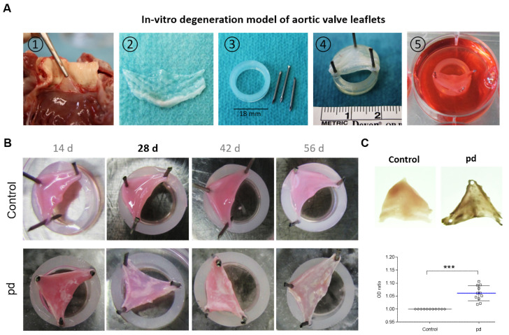
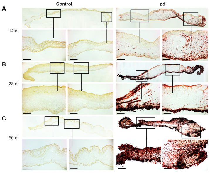
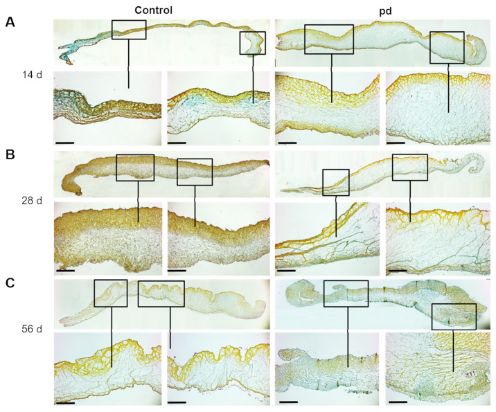
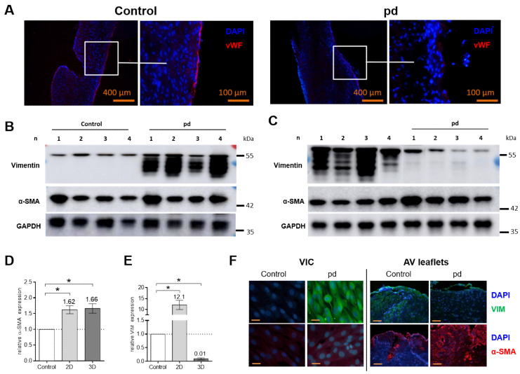
The hallmarks of calcific aortic valve disease (CAVD), an active and regulated process involving the creation of calcium nodules, lipoprotein accumulation, and chronic inflammation, are the significant changes that occur in the composition, organization, and mechanical properties of the extracellular matrix (ECM) of the aortic valve (AV). Most research regarding CAVD is based on experiments using two-dimensional (2D) cell culture or artificially created three-dimensional (3D) environments of valvular interstitial cells (VICs). Because the valvular ECM has a powerful influence in regulating pathological events, we developed an in vitro AV tissue culture model, which is more closely able to mimic natural conditions to study cellular responses underlying CAVD. AV leaflets, isolated from the hearts of 6-8-month-old sheep, were fixed with needles on silicon rubber rings to achieve passive tension and treated in vitro under pro-degenerative and pro-calcifying conditions. The degeneration of AV leaflets progressed over time, commencing with the first visible calcified domains after 14 d and winding up with the distinct formation of calcium nodules, heightened stiffness, and clear disruption of the ECM after 56 d. Both the expression of pro-degenerative genes and the myofibroblastic differentiation of VICs were altered in AV leaflets compared to that in VIC cultures. In this study, we have established an easily applicable, reproducible, and cost-effective in vitro AV tissue culture model to study pathological mechanisms underlying CAVD. The valvular ECM and realistic VIC-VEC interactions mimic natural conditions more closely than VIC cultures or 3D environments. The application of various culture conditions enables the examination of different pathological mechanisms underlying CAVD and could lead to a better understanding of the molecular mechanisms that lead to VIC degeneration and AS. Our model provides a valuable tool to study the complex pathobiology of CAVD and can be used to identify potential therapeutic targets for slowing disease progression.

Keywords: CAVD; aortic valve stenosis; calcific aortic valve disease; calcification; degeneration.


Conflict of interest statement

The authors declare no conflict of interest.

Figures

Figure 1
Figure 1
In vitro degeneration model of aortic valve leaflets. (A) Application of in vitro CAVD model: (1) Preparation of AV leaflets. (2) Excised AV leaflets after washing in PBS. (3) Required materials (silicon rubber rings and needles). (4) AV leaflet stretched on silicon rubber ring. (5) Cultivation of tensed AV leaflets. (B) Images of temporal progression of AV leaflet degeneration. White areas indicate calcified domains. (C) Representative transmitted light images of AV leaflets after 28 d cultivation and analysis of optical density (OD). Data (n = 8) are mean ± SEM. p-values are calculated by using Student’s t-test with Dunn’s multiple comparison post hoc test.; ***: p < 0.001. Pd, (pro-degenerative) condition.
Figure 2
Figure 2
Temporal progression of AV leaflet degeneration. Alizarin red S staining of AV leaflets under pro-degenerative (pd) conditions (β-GP + CaCl2) after 14 d (A), 28 d (B), and 56 d (C) compared to control conditions. Red indicates sites of biomineralization. Scale bar indicates 100 µm. Representative images of five different experiments are shown.
Figure 3
Figure 3
Temporal progression of ECM remodeling of AV leaflets. Movat’s pentachrome staining of AV leaflets under pro-degenerative (pd) conditions (β-GP + CaCl2) after 14 d (A), 28 d (B), and 56 d (C) compared to control conditions. Scale bar indicates 100 µm. Representative images of five different experiments are shown.
Figure 4
Figure 4
Analysis of endothelial layer and expression of VIM and α-SMA. (A) Immunohistological images with antibodies against von Willebrand factor (vWF) of AV leaflets under pro-degenerative (pd, β-GP + CaCl2) and control conditions after a 28 d cultivation period. Representative images of four different experiments are shown. Scale bar indicates 400 or 100 µm. Western blot images of 2D VIC cultures after 7 d (B) and AV leaflets after 28 d (C) for vimentin (VIM), smooth muscle alpha actin (α-SMA) and glyceraldehyde 3-phosphate dehydrogenase (GAPDH) under pd and control conditions. Density analysis for quantification of α-SMA (D) and VIM (E) in 2D VIC cultures and AV leaflets. Data (n = 4) are mean ± SEM. p-values are calculated by using Kruskal–Wallis test with Dunn’s multiple comparison post hoc test. *: p < 0.05. Data were normalized to GAPDH and expressed relative to control conditions. (F) Immunohistological images with antibodies against VIM (green) and α-SMA (red) of 2D VIC cultures and AV leaflets under pd and control conditions. Representative images of four different experiments are shown. DAPI, 4′,6-diamidino-2-phenylindole; VIC, Valvular interstitial cells.
Figure 5
Figure 5
Gene expression analysis. (A) Comparative RNA yields of 2D VIC cultures (7 d) and AV leaflets after 14, 28, and 56 d cultivation under pro-degenerative (pd) conditions (β-GP + CaCl2, dark column) compared to control conditions (white column). (B) Representative RNA integrity numbers (RINs) of AV leaflets after 28 d cultivation. (C) Analysis of gene expression of 2D VIC cultures (gray column, 7 d) and AV leaflets (dark column, 28 d) under pd conditions for alpha-1 type I collagen (Col1A1), alpha-1 type III collagen (Col3A1), alpha-1 type V collagen (Col5A1), transforming growth factor beta 1 (TGF-β1), vimentin (VIM), alpha smooth muscle actin (ACTA2), osteopontin (OPN), and osteoprotergerin (OPG) compared to control conditions (white column). Data (n = 6–8) are mean ± SEM. p-values are calculated by using Kruskal–Wallis test with Dunn’s multiple comparison post hoc test. *: p < 0.05; **: p < 0.01; ***: p < 0.001. FU, fluorescence units.
Figure 6
Figure 6
Comparison of pro-degenerative and pro-calcifying conditions. (A) Alizarin red S staining and quantification of 2D VIC cultures (n = 16) after 7 d under pro-degenerative (pd, β-GP + CaCl2; blue line) and pro-calcifying (pc, NaH2PO4, red line) conditions compared to control conditions. (B) Images and transmitted light images of AV leaflets after 28 d cultivation under pd and pc conditions and analysis of optical density (OD). Data (n = 6) are mean ± SEM. Alizarin red S (C), Movat’s pentachrome (D), and AP staining (E, purple areas) of AV leaflets under pd, pc, and control conditions after 28 d cultivation. Scale bar indicates 100 µm. Representative images of five different experiments are shown. Analysis of AP (F) and phosphate (G) in supernatants of 2D VIC cultures (d2–d7) and AV leaflet cultures (d3–d27) under pd (blue line) and pc (red line) conditions compared to control conditions (gray line). Data (n = 5) are mean ± SEM. p-values (*: pd vs. control, #: pc vs. control) are calculated by using Kruskal–Wallis test with Dunn’s multiple comparison post hoc test. * and #: p < 0.05; ** and ##: p < 0.01; *** and ###: p < 0.001.

References

    1. Adams H.S.L., Ashokkumar S., Newcomb A., MacIsaac A.I., Whitbourn R.J., Palmer S. Contemporary review of severe aortic stenosis. Intern. Med. J. 2019;49:297–305. doi: 10.1111/imj.14071. - DOI - PubMed
    1. Lindman B.R., Clavel M.A., Mathieu P., Iung B., Lancellotti P., Otto C.M., Pibarot P. Calcific aortic stenosis. Nat. Rev. Dis. Primers. 2016;2:16006. doi: 10.1038/nrdp.2016.6. - DOI - PMC - PubMed
    1. Perera S., Wijesinghe N., Ly E., Devlin G., Pasupati S. Outcomes of patients with untreated severe aortic stenosis in real-world practice. N. Z. Med. J. 2011;124:40–48. - PubMed
    1. Everett R.J., Clavel M.A., Pibarot P., Dweck M.R. Timing of intervention in aortic stenosis: A review of current and future strategies. Heart. 2018;104:2067–2076. doi: 10.1136/heartjnl-2017-312304. - DOI - PMC - PubMed
    1. Marquis-Gravel G., Redfors B., Leon M.B., Genereux P. Medical treatment of aortic stenosis. Circulation. 2016;134:1766–1784. doi: 10.1161/CIRCULATIONAHA.116.023997. - DOI - PubMed