Review
. 2021 Apr 26;11(5):393.
doi: 10.3390/life11050393.

Neurosurgical CSF Diversion in Idiopathic Intracranial Hypertension: A Narrative Review

Affiliations
Review

Neurosurgical CSF Diversion in Idiopathic Intracranial Hypertension: A Narrative Review

Geraint J Sunderland et al. Life (Basel). .

Abstract

The prevalence of idiopathic intracranial hypertension (IIH), a complex disorder, is increasing globally in association with obesity. The IIH syndrome occurs as the result of elevated intracranial pressure, which can cause permanent visual impairment and loss if not adequately managed. CSF diversion via ventriculoperitoneal and lumboperitoneal shunts is a well-established strategy to protect vision in medically refractory cases. Success of CSF diversion is compromised by high rates of complication; including over-drainage, obstruction, and infection. This review outlines currently used techniques and technologies in the management of IIH. Neurosurgical CSF diversion is a vital component of the multidisciplinary management of IIH.

Keywords: anti-siphon device; cerebrospinal fluid; idiopathic intracranial hypertension; lumboperitoneal shunt; neurosurgery; programmable valve; pseudotumour cerebri; ventriculoperitoneal shunt.


Conflict of interest statement

The authors declare no conflict of interest.

Figures

Figure 1
Figure 1
Illustrative case of a 9-year old boy presenting with chronic headaches and visual disturbance. Examination revealed bilateral papilloedema. The initial CT did not demonstrate a pathological mass lesion, but did show characteristic small ventricles (A). Lumbar puncture was equivocal with an opening pressure of 25 cmH2O. Pre-shunt invasive ICP monitoring demonstrated characteristic pathological plateau waveforms (B). A VP shunt was placed under EM guidance. Post-operative axial (C) and coronal (D) CT demonstrated satisfactory position of the ventricular catheter within the frontal horn of the left lateral ventricle.
Figure 2
Figure 2
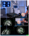
Demonstration of electromagnetic VP shunt insertion setup using the Medtronic AxiEM™ system. Screenshots from Medtronic promotional video.
Figure 3
Figure 3
Algorithm outlining a systematic approach to assessment and management of IIH patients with VP shunts presenting with an exacerbation of headache symptoms. Adapted from Mollan et al 2018 [12]., used under creative commons CC BY 4.0 license.
Figure 4
Figure 4
Density histogram demonstrating the age distribution of IIH patients compared with non-IIH cohorts (aged 5–65 years) from the BASICS trial.

References

    1. Durcan F.J., Corbett J.J., Wall M. The Incidence of Pseudotumor Cerebri. Arch. Neurol. 1988;45:875–877. doi: 10.1001/archneur.1988.00520320065016. - DOI - PubMed
    1. Ball A.K., Clarke C.E. Idiopathic intracranial hypertension. Lancet Neurol. 2006;5:433–442. doi: 10.1016/S1474-4422(06)70442-2. - DOI - PubMed
    1. Kilgore K.P., Lee M.S., Leavitt J.A., Mokri B., Hodge D.O., Frank R.D., Chen J.J. Re-evaluating the Incidence of Idiopathic Intracranial Hypertension in an Era of Increasing Obesity. Ophthalmology. 2017;124:697–700. doi: 10.1016/j.ophtha.2017.01.006. - DOI - PMC - PubMed
    1. Curry W.T., Butler W.E., Barker F.G. Rapidly Rising Incidence of Cerebrospinal Fluid Shunting Procedures for Idiopathic Intracranial Hypertension in the United States, 1988–2002. Neurosurgery. 2005;57:97–108. doi: 10.1227/01.NEU.0000163094.23923.E5. - DOI - PubMed
    1. Mollan S.P., Aguiar M., Evison F., Frew E., Sinclair A.J. The expanding burden of idiopathic intracranial hypertension. Eye. 2019;33:478–485. doi: 10.1038/s41433-018-0238-5. - DOI - PMC - PubMed

LinkOut - more resources