Review
. 2021 Apr 26;13(9):2093.
doi: 10.3390/cancers13092093.

Use of PET Imaging in Neuro-Oncological Surgery

Affiliations
Review

Use of PET Imaging in Neuro-Oncological Surgery

Adrien Holzgreve et al. Cancers (Basel). .

Abstract

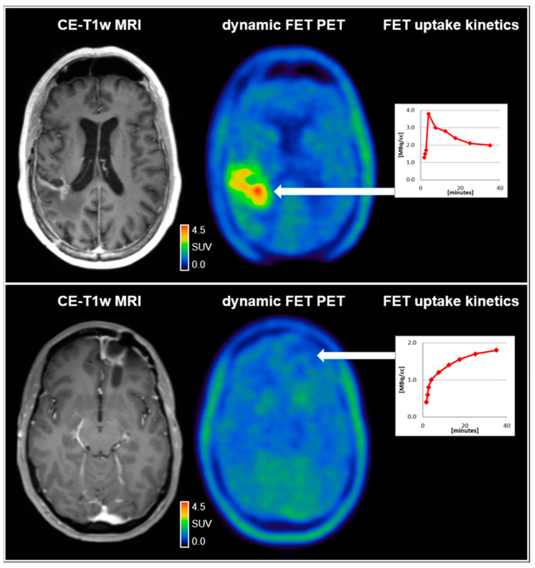
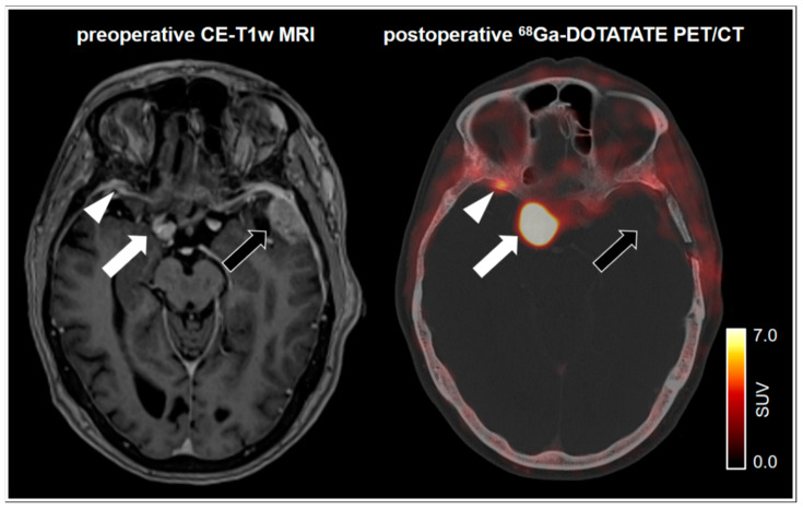
This review provides an overview of current applications and perspectives of PET imaging in neuro-oncological surgery. The past and future of PET imaging in the management of patients with glioma and brain metastases are elucidated with an emphasis on amino acid tracers, such as O-(2-[18F]fluoroethyl)-L-tyrosine (18F-FET). The thematic scope includes surgical resection planning, prognostication, non-invasive prediction of molecular tumor characteristics, depiction of intratumoral heterogeneity, response assessment, differentiation between tumor progression and treatment-related changes, and emerging new tracers. Furthermore, the role of PET using specific somatostatin receptor ligands for the management of patients with meningioma is discussed. Further advances in neuro-oncological imaging can be expected from promising new techniques, such as hybrid PET/MR scanners and the implementation of artificial intelligence methods, such as radiomics.

Keywords: FET PET; PET imaging; brain metastasis; glioblastoma; glioma; meningioma; neuro-oncological surgery; neurosurgery; somatostatin receptor.


Conflict of interest statement

The authors declare no conflict of interest.

Figures

Figure 1
Figure 1
(upper row) A 59-year-old male patient diagnosed with an IDH-wild-type glioblastoma (WHO CNS grade 4). Following resection and chemoradiation with temozolomide, the contrast-enhanced MRI (CE-T1w MRI) suggested tumor relapse in the right parietal region 7 months after completing radiotherapy. Accordingly, the dynamic FET PET scan revealed pathologically increased FET uptake right parietal (TBRmax, 4.2) and decreased time–activity curve; (lower row). A 37-year-old female patient diagnosed with an IDH-wild-type glioblastoma (WHO CNS grade 4). Following resection and chemoradiation with temozolomide, the contrast-enhanced MRI suggested tumor relapse in the left frontal region 7 months after completing radiotherapy. In contrast to the patient in the upper row, the FET uptake in the left frontal region was not pathologically increased (TBRmax, 1.6) with a steadily increasing time–activity curve, indicating reactive treatment-related changes. SUV = standardized uptake value.
Figure 2
Figure 2
75-year-old female patient with a left frontotemporal transitional meningioma of the WHO grade 1. 68Ga-DOTATATE PET/CT shows no postoperative remnants of the left frontotemporal tumor (black arrow). Pronounced tracer uptake in the right parasellar region indicates a meningioma in correlation to the MRI (white arrow). Notably, a small focal uptake posterior to the right orbital region indicates an additional meningioma. In spatial correspondence, the MRI shows equivocal findings (white arrowhead). SUV = standardized uptake value.

References

    1. Gambhir S.S. Molecular imaging of cancer with positron emission tomography. Nat. Rev. Cancer. 2002;2:683–693. doi: 10.1038/nrc882. - DOI - PubMed
    1. La Fougère C., Suchorska B., Bartenstein P., Kreth F.-W., Tonn J.-C. Molecular imaging of gliomas with PET: Opportunities and limitations. Neuro-Oncology. 2011;13:806–819. doi: 10.1093/neuonc/nor054. - DOI - PMC - PubMed
    1. Bergström M., Collins V.P., Ehrin E., Ericson K., Eriksson L., Greitz T., Halldin C., Von Hoist H., Långström B., Lilja A., et al. Discrepancies in Brain Tumor Extent as Shown by Computed Tomography and Positron Emission Tomography Using [68Ga]EDTA, [11C]Glucose, and [11C]Methionine. J. Comput. Assist. Tomogr. 1983;7:1062–1066. doi: 10.1097/00004728-198312000-00022. - DOI - PubMed
    1. Karlberg A., Berntsen E.M., Johansen H., Skjulsvik A.J., Reinertsen I., Dai H.Y., Xiao Y., Rivaz H., Borghammer P., Solheim O., et al. 18F-FACBC PET/MRI in Diagnostic Assessment and Neurosurgery of Gliomas. Clin. Nucl. Med. 2019;44:550–559. doi: 10.1097/RLU.0000000000002610. - DOI - PubMed
    1. Law I., Albert N.L., Arbizu J., Boellaard R., Drzezga A., Galldiks N., La Fougère C., Langen K.-J., Lopci E., Lowe V., et al. Joint EANM/EANO/RANO practice guidelines/SNMMI procedure standards for imaging of gliomas using PET with radiolabelled amino acids and [18F]FDG: Version 1.0. Eur. J. Nucl. Med. Mol. Imaging. 2019;46:540–557. doi: 10.1007/s00259-018-4207-9. - DOI - PMC - PubMed

LinkOut - more resources