

Zoledronic acid modulates osteoclast apoptosis through activation of the NF-κB signaling pathway in ovariectomized rats
- PMID: 33926259
- PMCID: PMC8719043
- DOI: 10.1177/15353702211011052
Zoledronic acid modulates osteoclast apoptosis through activation of the NF-κB signaling pathway in ovariectomized rats
Abstract
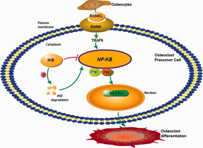
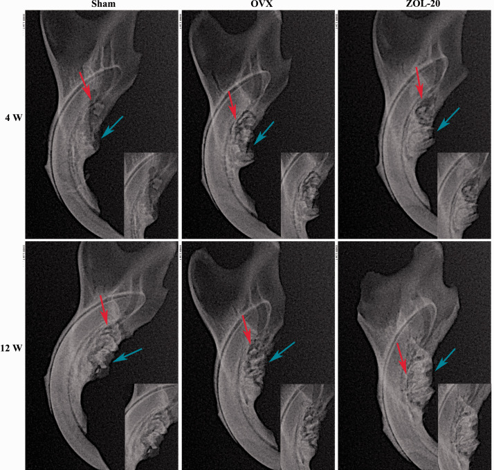
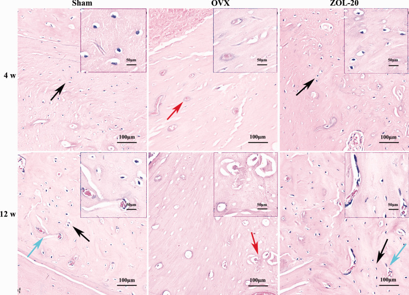
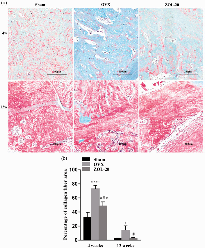
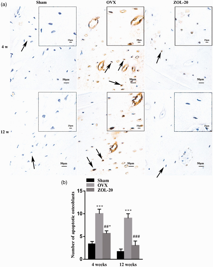
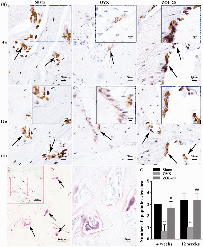
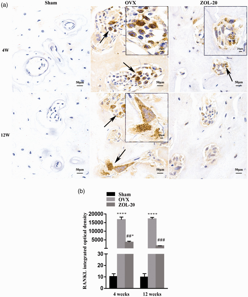
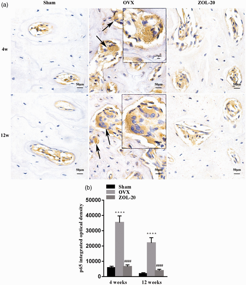
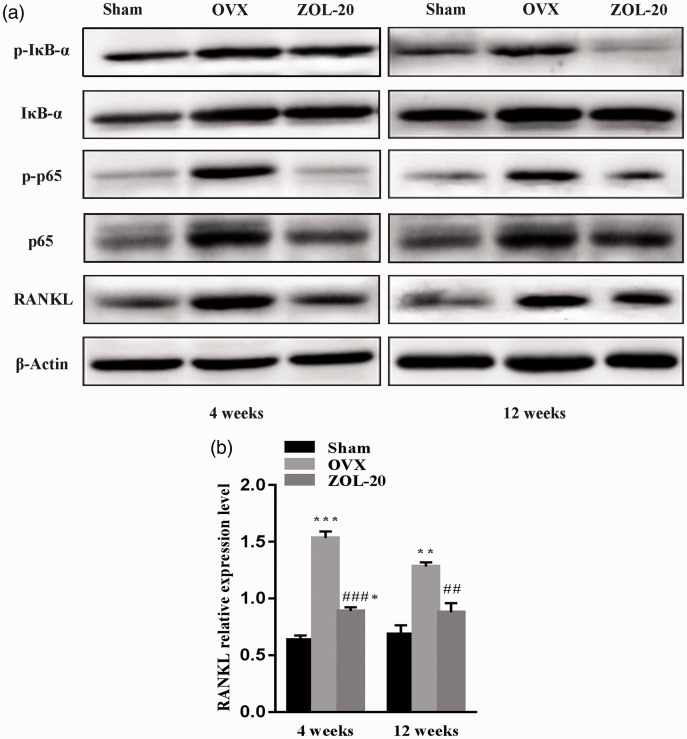
Bone mass loss (osteoporosis) seen in postmenopausal women is an adverse factor for implant denture. Using an ovariectomized rat model, we studied the mechanism of estrogen-deficiency-caused bone loss and the therapeutic effect of Zoledronic acid. We observed that ovariectomized-caused resorption of bone tissue in the mandible was evident at four weeks and had not fully recovered by 12 weeks post-ovariectomized compared with the sham-operated controls. Further evaluation with a TUNEL assay showed ovariectomized enhanced apoptosis of osteoblasts but inhibited apoptosis of osteoclasts in the mandible. Zoledronic acid given subcutaneously as a single low dose was shown to counteract both of these ovariectomized effects. Immunohistochemical staining showed that ovariectomized induced the protein levels of RANKL and the 65-kD subunit of the NF-κB complex mainly in osteoclasts, as confirmed by staining for TRAP, a marker for osteoclasts, whereas zoledronic acid inhibited these inductions. Western blotting showed that the levels of RANKL, p65, as well as the phosphorylated form of p65, and IκB-α were all higher in the ovariectomized group than in the sham and ovariectomized + zoledronic acid groups at both the 4th- and 12th-week time points in the mandible. These data collectively suggest that ovariectomized causes bone mass loss by enhancing apoptosis of osteoblasts and inhibiting apoptosis of osteoclasts. In osteoclasts, these cellular effects may be achieved by activating RANKL-NF-κB signalling. Moreover, zoledronic acid elicits its therapeutic effects in the mandible by counteracting these cellular and molecular consequences of ovariectomized.
Keywords: NF-κB signaling pathway; RANKL; Zoledronic acid; osteoblast; osteoclast; osteoporosis.
Conflict of interest statement
Figures
Similar articles
-
Glaucocalyxin A suppresses osteoclastogenesis induced by RANKL and osteoporosis induced by ovariectomy by inhibiting the NF-κB and Akt pathways.J Ethnopharmacol. 2021 Aug 10;276:114176. doi: 10.1016/j.jep.2021.114176. Epub 2021 Apr 30. J Ethnopharmacol. 2021. PMID: 33933570
-
Hydrogen gas protects against ovariectomy-induced osteoporosis by inhibiting NF-κB activation.Menopause. 2019 Jul;26(7):785-792. doi: 10.1097/GME.0000000000001310. Menopause. 2019. PMID: 31083022
-
Longan fruit increase bone mineral density in zebrafish and ovariectomized rat by suppressing RANKL-induced osteoclast differentiation.Phytomedicine. 2019 Jun;59:152910. doi: 10.1016/j.phymed.2019.152910. Epub 2019 Apr 2. Phytomedicine. 2019. PMID: 30978650
-
α-Linolenic Acid Inhibits Receptor Activator of NF-κB Ligand Induced (RANKL-Induced) Osteoclastogenesis and Prevents Inflammatory Bone Loss via Downregulation of Nuclear Factor-KappaB-Inducible Nitric Oxide Synthases (NF-κB-iNOS) Signaling Pathways.Med Sci Monit. 2017 Oct 24;23:5056-5069. doi: 10.12659/msm.904795. Med Sci Monit. 2017. PMID: 29061958 Free PMC article.
-
Research advances on silence information regulator 6 as a potential therapeutic target for bone regeneration and repair.Zhejiang Da Xue Xue Bao Yi Xue Ban. 2024 Aug 25;53(4):427-433. doi: 10.3724/zdxbyxb-2023-0615. Zhejiang Da Xue Xue Bao Yi Xue Ban. 2024. PMID: 39183069 Free PMC article. Review. Chinese, English.
Cited by
-
Increased bone mass but delayed mineralization: in vivo and in vitro study for zoledronate in bone regeneration.BMC Oral Health. 2024 Sep 27;24(1):1146. doi: 10.1186/s12903-024-04906-2. BMC Oral Health. 2024. PMID: 39334089 Free PMC article.
-
Oestradiol Contributes to Differential Antitumour Effects of Adjuvant Zoledronic Acid Observed Between Pre- and Post-Menopausal Women.Front Endocrinol (Lausanne). 2021 Oct 18;12:749428. doi: 10.3389/fendo.2021.749428. eCollection 2021. Front Endocrinol (Lausanne). 2021. PMID: 34733240 Free PMC article.
-
Mild antiresorptive activity of an anti-vascular endothelial growth factor A antibody and sunitinib in a rat model of bone resorption.Bone Rep. 2025 Mar 17;25:101837. doi: 10.1016/j.bonr.2025.101837. eCollection 2025 Jun. Bone Rep. 2025. PMID: 40177629 Free PMC article.
-
A Systematic Review of the Effects of Bisphosphonates on Osteoblasts In Vitro.Calcif Tissue Int. 2025 Jun 16;116(1):86. doi: 10.1007/s00223-025-01390-w. Calcif Tissue Int. 2025. PMID: 40523987 Free PMC article.
-
METTL7A-mediated m6A modification of corin reverses bisphosphonates-impaired osteogenic differentiation of orofacial BMSCs.Int J Oral Sci. 2024 May 23;16(1):42. doi: 10.1038/s41368-024-00303-1. Int J Oral Sci. 2024. PMID: 38782892 Free PMC article.
References
-
- Buser D, Sennerby L, De Bruyn H. Modern implant dentistry based on osseointegration: 50 years of progress, current trends and open questions. Periodontol 2000 2017; 73:7–21 - PubMed
-
- Charatchaiwanna A, Rojsiraphisa T, Aunmeungtong W, Reichart PA, Khongkhunthian P. Mathematical equations for dental implant stability patterns during the osseointegration period, based on previous resonance frequency analysis studies. Clin Implant Dent Relat Res 2019; 21:1028–40 - PubMed
-
- de Medeiros F, Kudo G, Leme BG, Saraiva PP, Verri FR, Honorio HM, Pellizzer EP, Santiago JJ. Dental implants in patients with osteoporosis: a systematic review with meta-analysis. Int J Oral Maxillofac Surg 2018; 47:480–91 - PubMed
Publication types
MeSH terms
Substances
LinkOut - more resources
Full Text Sources
Other Literature Sources