. 2021 Apr 29;13(1):97.
doi: 10.1186/s13148-021-01080-y.

Characteristics of epigenetic aging across gestational and perinatal tissues

Affiliations

Characteristics of epigenetic aging across gestational and perinatal tissues

Linda Dieckmann et al. Clin Epigenetics. .

Abstract

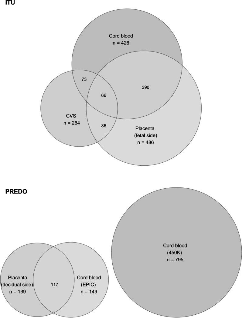
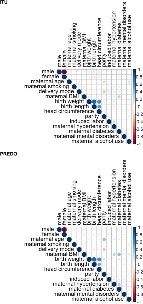
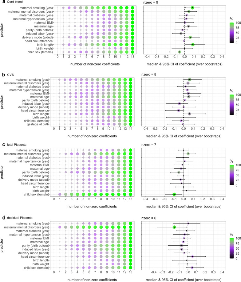
Background: Epigenetic clocks have been used to indicate differences in biological states between individuals of same chronological age. However, so far, only few studies have examined epigenetic aging in newborns-especially regarding different gestational or perinatal tissues. In this study, we investigated which birth- and pregnancy-related variables are most important in predicting gestational epigenetic age acceleration or deceleration (i.e., the deviation between gestational epigenetic age estimated from the DNA methylome and chronological gestational age) in chorionic villus, placenta and cord blood tissues from two independent study cohorts (ITU, n = 639 and PREDO, n = 966). We further characterized the correspondence of epigenetic age deviations between these tissues.

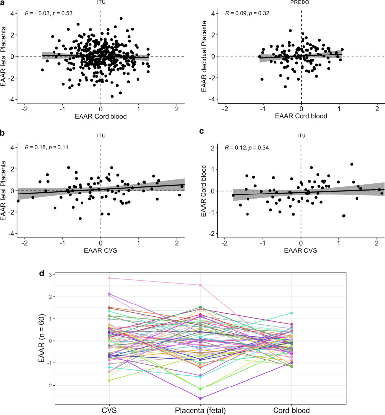
Results: Among the most predictive factors of epigenetic age deviations in single tissues were child sex, birth length, maternal smoking during pregnancy, maternal mental disorders until childbirth, delivery mode and parity. However, the specific factors related to epigenetic age deviation and the direction of association differed across tissues. In individuals with samples available from more than one tissue, relative epigenetic age deviations were not correlated across tissues.

Conclusion: Gestational epigenetic age acceleration or deceleration was not related to more favorable or unfavorable factors in one direction in the investigated tissues, and the relative epigenetic age differed between tissues of the same person. This indicates that epigenetic age deviations associate with distinct, tissue specific, factors during the gestational and perinatal period. Our findings suggest that the epigenetic age of the newborn should be seen as a characteristic of a specific tissue, and less as a general characteristic of the child itself.

Keywords: Chorionic villi; Cord blood; Early development; Epigenetic age; Epigenetic clocks; Perinatal tissues; Placenta.


Conflict of interest statement

EB is the coinventor of FKBP5: a novel target for antidepressant therapy, European Patent no. EP 1687443 B1, and receives a research grant from Böhringer Ingelheim for a collaboration on functional investigations of FKBP5. Otherwise, the authors declare that they have no competing interests.

Figures

Fig. 1
Fig. 1
Sample overview for both cohorts used. Samples with methylation data available from different tissues in ITU and PREDO. In total, the ITU data set comprised 693 individuals after QC, with 264 CVS, 486 fetal placenta and 426 cord blood samples. For some individuals, samples were available from several tissues, indicated by overlapping circles. The final PREDO data set comprised 171 individuals after QC processed with the EPIC array, and additional 795 individuals processed with the 450 K array. From the EPIC data, 139 samples were available from placenta, and 149 samples from cord blood. The number of individuals with data from both tissues is again illustrated by the overlapping circles
Fig. 2
Fig. 2
Pearson correlations among the predictor variables for ITU (N = 693) and PREDO (N = 171)
Fig. 3
Fig. 3
Outcomes of elastic net regression models in different tissues. Associations between birth- and pregnancy-related variables (predictors) and EAAR (adjusted for gestational age at time of sampling, cell types and ancestry-related information). Depicted are the percentages of variable occurrence in bootstrap models with different number of non-zero coefficients (left) and the coefficients of variables in the final model (right) in cord blood from ITU (a), CVS from ITU (b), fetal placenta from ITU (c) and in decidual placenta from PREDO (d). The color coding shows the percentage of occurrence of a variable in the model over bootstraps and the size of the circle is proportional
Fig. 4
Fig. 4
Relationship of epigenetic age acceleration/deceleration between different tissues. In children with more than one tissue available, the relationship of epigenetic age acceleration or deceleration between the respective tissues can be illustrated. Depicted are the scatter plots of EAAR for (a) cord blood and placenta from both ITU (n = 363) and PREDO (n = 116), (b) CVS and placenta from ITU (n = 78), and (c) CVS and cord blood from ITU (n = 66). The regression line is plotted together with a 95% confidence interval, and the Pearson correlation coefficient is depicted. Individual differences in EAARs between CVS, placenta and cord blood from ITU are further illustrated (d) for n = 60 children from ITU, where each color represents one individual.

References

    1. Jones MJ, Goodman SJ, Kobor MS. DNA methylation and healthy human aging. Aging Cell. 2015;14(6):924–932. doi: 10.1111/acel.12349. - DOI - PMC - PubMed
    1. Xiao FH, Wang HT, Kong QP. Dynamic DNA methylation during aging: a "prophet" of age-related outcomes. Front Genet. 2019;10:107. doi: 10.3389/fgene.2019.00107. - DOI - PMC - PubMed
    1. Field AE, Robertson NA, Wang T, Havas A, Ideker T, Adams PD. DNA methylation clocks in aging: categories, causes, and consequences. Mol Cell. 2018;71(6):882–895. doi: 10.1016/j.molcel.2018.08.008. - DOI - PMC - PubMed
    1. Horvath S. DNA methylation age of human tissues and cell types. Genome Biol. 2013;14. - PMC - PubMed
    1. Hannum G, Guinney J, Zhao L, Zhang L, Hughes G, Sadda S, et al. Genome-wide methylation profiles reveal quantitative views of human aging rates. Mol Cell. 2013;49(2):359–367. doi: 10.1016/j.molcel.2012.10.016. - DOI - PMC - PubMed

Publication types