. 2021 Oct;11(10):2488-2505.
doi: 10.1158/2159-8290.CD-20-1669. Epub 2021 Apr 29.

FGFR2 Extracellular Domain In-Frame Deletions Are Therapeutically Targetable Genomic Alterations That Function as Oncogenic Drivers in Cholangiocarcinoma

Affiliations

FGFR2 Extracellular Domain In-Frame Deletions Are Therapeutically Targetable Genomic Alterations That Function as Oncogenic Drivers in Cholangiocarcinoma

James M Cleary et al. Cancer Discov. 2021 Oct.

Abstract

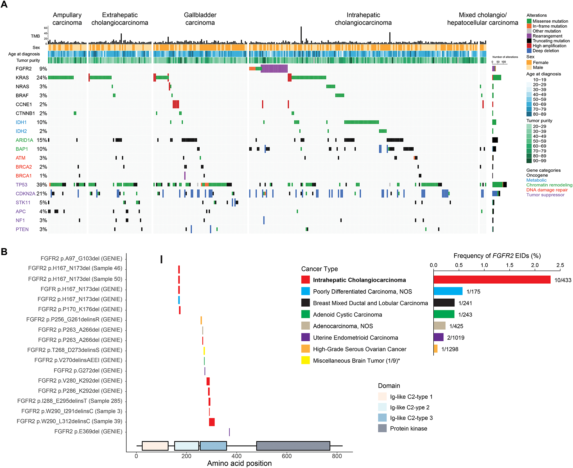
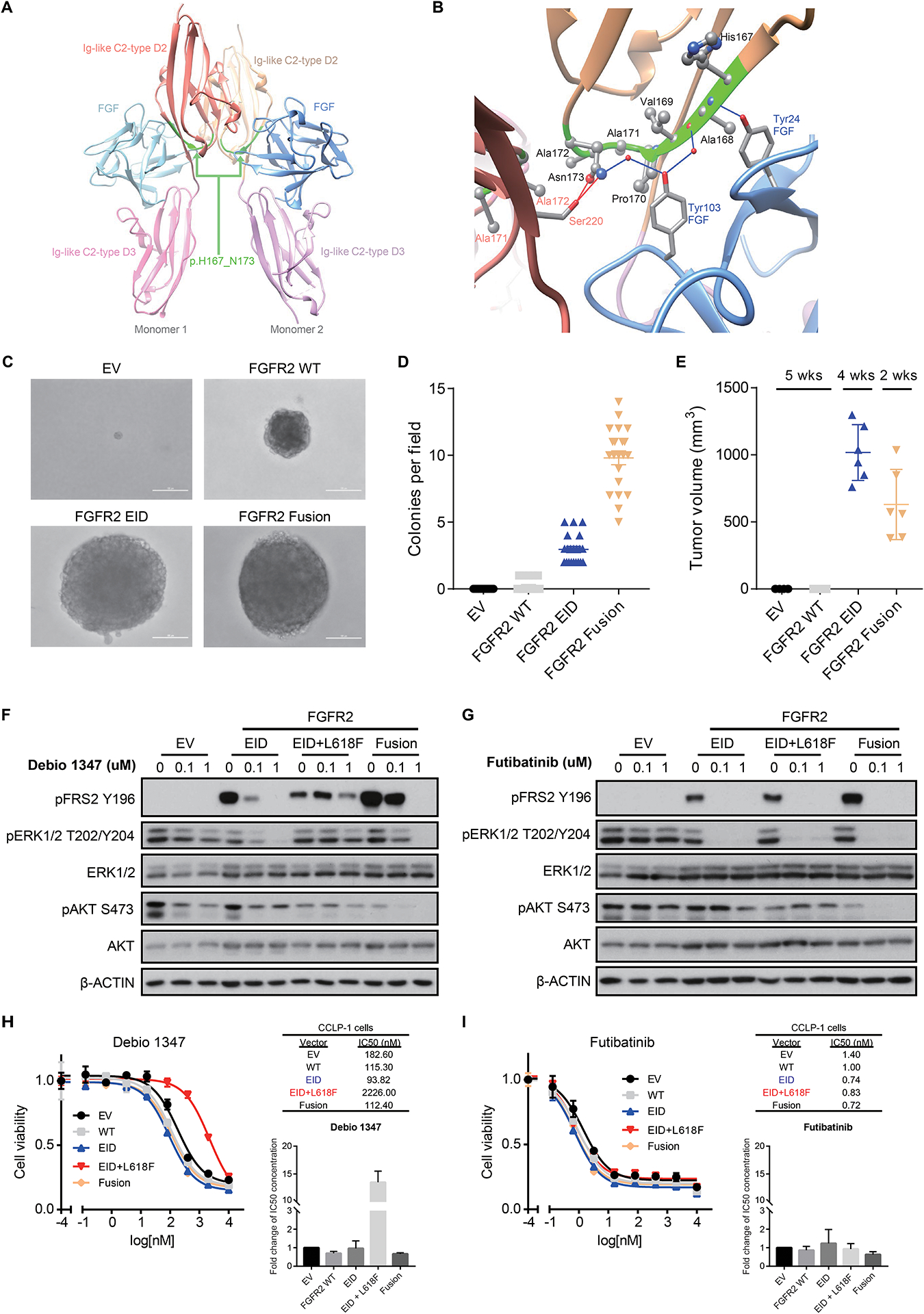
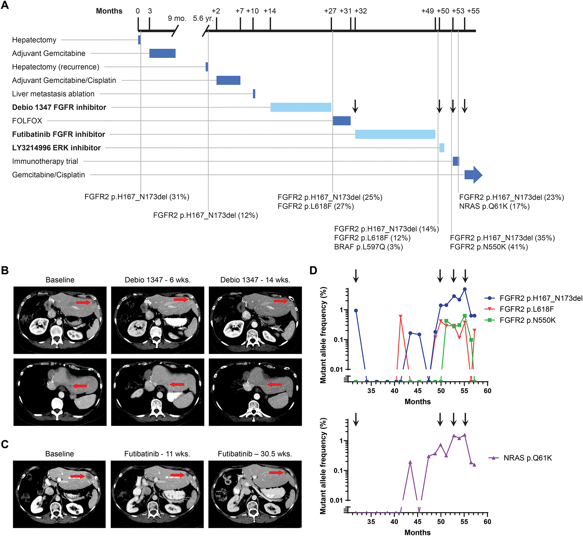
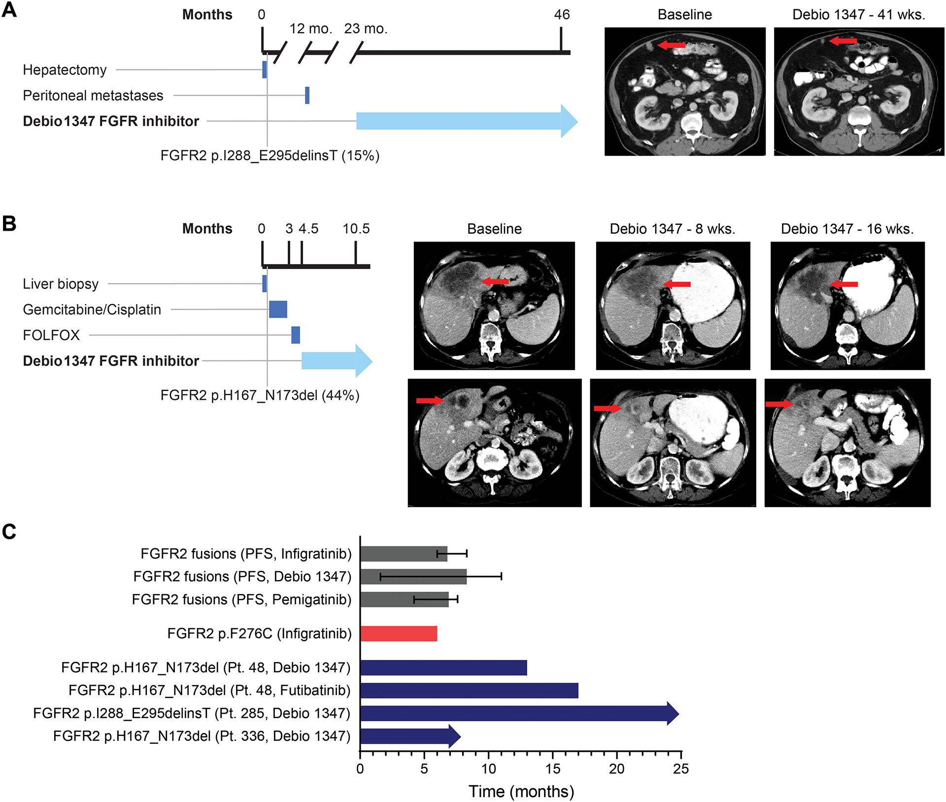
We conducted next-generation DNA sequencing on 335 biliary tract cancers and characterized the genomic landscape by anatomic site within the biliary tree. In addition to frequent FGFR2 fusions among patients with intrahepatic cholangiocarcinoma (IHCC), we identified FGFR2 extracellular domain in-frame deletions (EID) in 5 of 178 (2.8%) patients with IHCC, including two patients with FGFR2 p.H167_N173del. Expression of this FGFR2 EID in NIH3T3 cells resulted in constitutive FGFR2 activation, oncogenic transformation, and sensitivity to FGFR inhibitors. Three patients with FGFR2 EIDs were treated with Debio 1347, an oral FGFR1/2/3 inhibitor, and all showed partial responses. One patient developed an acquired L618F FGFR2 kinase domain mutation at disease progression and experienced a further partial response for 17 months to an irreversible FGFR2 inhibitor, futibatinib. Together, these findings reveal FGFR2 EIDs as an alternative mechanism of FGFR2 activation in IHCC that predicts sensitivity to FGFR inhibitors in the clinic. SIGNIFICANCE: FGFR2 EIDs are transforming genomic alterations that occur predominantly in patients with IHCC. These FGFR2 EIDs are sensitive to FGFR inhibition in vitro, and patients with these alterations benefited from treatment with FGFR inhibitors in the clinic.This article is highlighted in the In This Issue feature, p. 2355.


Figures

Figure 1:
Figure 1:. Genomic alterations in 335 biliary tract cancers identified by targeted next generation sequencing.
A. Co-mutation plot highlighting frequently altered genes in biliary tract cancers. Columns represent individual patients with biliary tract cancer, and rows indicate somatic genomic alterations. Each sample’s mutation per megabase (TMB) is represented by a histogram. The types of genomic alterations are color coded. Genes are listed according to the following functional classes: oncogenes, metabolic chromatin remodeling, DNA damage repair, and tumor suppressors (from top to bottom, color coded). B. Schematic depiction of the intragenic location of extracellular domain FGFR2 EIDs and their alteration frequencies by tumor type in the GENIE database. FGFR2 EIDs are enriched in samples from IHCC patients. * indicates that the “Miscellaneous brain tumor” cancer type was not included in the frequency graph due to low sample number.
Figure 2:
Figure 2:. FGFR2 EIDs are oncogenic and confer sensitivity to FGFR inhibitors.
A, B. Modeling alteration p.H167_N173del within the three-dimensional structure of FGFR2. Panel A shows a global view of the p.H167_N173 deletion (ribbon highlighted in green) located in both Ig-like C2-type D2 extracellular domains of FGFR2 dimeric structure; refer to Supplemental Figure 7 (deletion #1) for mapping onto the protein sequence. As shown more closely in panel B, p.H167_N173 (ribbon highlighted in green) is an extended deletion, mostly included in a beta-strand of the C2-type D2 domain (ribbon in beige), and makes several intermolecular interactions with both the FGF ligand (ribbon and residue names in blue) and the C2-type D2 domain of the other monomer (ribbon and residue names in red). Intermolecular hydrogen-bonds (direct or water-mediated) between p.H167_N173 residues and FGF or the other monomer are shown as blue and red lines, respectively. Van der Waals contacts are not displayed explicitly. C, D. Expression of FGFR2 p.H167_N173del in NIH3T3 cells is sufficient for transformation in soft agar colony formation assays. Cells were transduced with retroviral vectors containing empty vector control (EV), wild-type FGFR2 (FGFR2 WT), FGFR2 p.H167_N173del (FGFR2 EID), or FGFR2-OPTN fusion (FGFR2 Fusion). Representative images (C) and quantification of number of colonies per field of view (D) are shown. Line and bars indicate mean with standard error, with 21 fields of view assessed across three biologic replicates. E. Volume of subcutaneous tumors forming in NSG mice following implantation of NIH3T3 cells expressing the indicated constructs (n=6 mice per condition). Both the FGFR2 p.H167_N173del EID and the FGFR2-PHGDH fusion (FGFR2 Fusion) induce tumor formation. Expression of the FGFR2-PHGDH fusion led to slightly faster tumor growth necessitating euthanasia at an earlier time point. Line and bars indicate mean with standard deviation. F, G. Expression of FGFR2 p.H167_N173del (EID), the EID with an L618F mutant kinase domain (EID+L618F), and FGFR2-PHGDH fusions (Fusion) lead to constitutive FGFR signaling (induction of FRS2) in NIH3T3 cells. FGFR2 EIDs and fusions are sensitive to treatment with Debio1347 (reduction in pFRS2 and pERK), whereas the L618F mutation causes resistance (F); all three of these FGFR2 alterations are inhibited by futibatinib (TAS-120) (G). H, I. Cell viability assays in CCLP-1 cells harboring empty vector control (EV), wild-type FGFR2 (WT), FGFR2 p.H167_N173del (EID), the co-occurring FGFR2 kinase mutation L618F (EID+L618F), or FGFR2-PHGDH fusion (Fusion) and treated with Debio 1347 (H) or futibatinib (I). Drug response measurements were performed in three independent experiments with each consisting of two technical replicates. Inset graphs demonstrate the average fold change in IC50 between conditions. Representative dose response curves and IC50 values are shown from a single experiment, with error bars on the curves representing standard deviation from 2 technical replicates.
Figure 3:
Figure 3:. Treatment course for Patient 46 with intrahepatic cholangiocarcinoma and FGFR2 p.H167_N173del EID.
A. Timeline depicts patient’s treatment course and duration of therapy. Notable somatic mutations detected on liver biopsies (black vertical lines) are listed below the timeline. The mutation allele fraction (MAF) is listed as a percentage next to the genomic alteration. Arrows correspond to time points when a new treatment was started, in reference to droplet digital PCR analysis in panel D. Treatments with FGFR and MAPK targeted agents are highlighted in bold and duration delineated with a lighter blue color. B. Computed tomography (CT) scans demonstrating the patient’s liver lesions at baseline and after 6 and 14 weeks of Debio 1347 treatment. C. CT scans evaluating the patient’s liver lesions at baseline and after 11 and 30.5 weeks of futibatinib (TAS-120) treatment. D. Selected alterations identified retrospectively in circulating cell free DNA by droplet digital PCR of serially banked plasma samples. Arrows correspond to time points when a new treatment was started in the clinical timeline in panel A.
Figure 4:
Figure 4:. Patients with FGFR2 EIDs have prolonged clinical responses to FGFR inhibitors.
A. Timeline and computed tomography scans demonstrating Patient 285’s peritoneal lesion at baseline and after 41 weeks of Debio 1347 treatment. B. Timeline and computed tomography scans demonstrating Patient 336’s liver mass at baseline and after 8 and 16 weeks of treatment with Debio 1347. C. Bar graph depicting progression-free survival (PFS) of patients with cholangiocarcinoma harboring an FGFR2 fusion treated with infigratinib (17), Debio 1347, and pemigatinib (3). Error bars indicate 95% confidence intervals. Graph also shows time on treatment for a cholangiocarcinoma patient with an FGFR2 F276C extracellular domain mutation who was treated with infigratinib (41), and time on treatment for three IHCC FGFR2 EID patients described in this manuscript. Arrows indicate that treatment is ongoing.

References

    1. Rizvi S, Khan SA, Hallemeier CL, Kelley RK, Gores GJ. Cholangiocarcinoma - evolving concepts and therapeutic strategies. Nat Rev Clin Oncol 2018;15(2):95–111 doi 10.1038/nrclinonc.2017.157. - DOI - PMC - PubMed
    1. Valle JW, Lamarca A, Goyal L, Barriuso J, Zhu AX. New Horizons for Precision Medicine in Biliary Tract Cancers. Cancer Discov 2017;7(9):943–62 doi 10.1158/2159-8290.Cd-17-0245. - DOI - PMC - PubMed
    1. Abou-Alfa GK, Sahai V, Hollebecque A, Vaccaro G, Melisi D, Al-Rajabi R, et al. Pemigatinib for previously treated, locally advanced or metastatic cholangiocarcinoma: a multicentre, open-label, phase 2 study. Lancet Oncol 2020;21(5):671–84 doi 10.1016/s1470-2045(20)30109-1. - DOI - PMC - PubMed
    1. Javle M, Lowery M, Shroff RT, Weiss KH, Springfeld C, Borad MJ, et al. Phase II Study of BGJ398 in Patients With FGFR-Altered Advanced Cholangiocarcinoma. J Clin Oncol 2018;36(3):276–82 doi 10.1200/JCO.2017.75.5009. - DOI - PMC - PubMed
    1. Goyal L, Shi L, Liu LY, Fece de la Cruz F, Lennerz JK, Raghavan S, et al. TAS-120 Overcomes Resistance to ATP-Competitive FGFR Inhibitors in Patients with FGFR2 Fusion-Positive Intrahepatic Cholangiocarcinoma. Cancer Discov 2019;9(8):1064–79 doi 10.1158/2159-8290.CD-19-0182. - DOI - PMC - PubMed

Publication types

MeSH terms

Substances