. 2021 Apr 30;372(6541):eabb8699.
doi: 10.1126/science.abb8699.

Stepwise pathogenic evolution of Mycobacterium abscessus

Affiliations

Stepwise pathogenic evolution of Mycobacterium abscessus

Josephine M Bryant et al. Science. .

Abstract

Although almost all mycobacterial species are saprophytic environmental organisms, a few, such as Mycobacterium tuberculosis, have evolved to cause transmissible human infection. By analyzing the recent emergence and spread of the environmental organism M. abscessus through the global cystic fibrosis population, we have defined key, generalizable steps involved in the pathogenic evolution of mycobacteria. We show that epigenetic modifiers, acquired through horizontal gene transfer, cause saltational increases in the pathogenic potential of specific environmental clones. Allopatric parallel evolution during chronic lung infection then promotes rapid increases in virulence through mutations in a discrete gene network; these mutations enhance growth within macrophages but impair fomite survival. As a consequence, we observe constrained pathogenic evolution while person-to-person transmission remains indirect, but postulate accelerated pathogenic adaptation once direct transmission is possible, as observed for M. tuberculosis Our findings indicate how key interventions, such as early treatment and cross-infection control, might restrict the spread of existing mycobacterial pathogens and prevent new, emergent ones.


Conflict of interest statement

Competing interests: none.

Figures

Figure 1
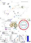
Figure 1. Saltational evolution of M. abscessus dominant circulating clones.
(A) Pangenome graph of M. abscessus (constructed using Panaroo [15]), where nodes represent clusters of orthologous genes and two nodes are connected by an edge if they are adjacent on a contig in any sample from the population, defines gene gain events associated with the emergence of DCC1 (purple), DCC2 (blue), and DCC3 (orange). For illustration purposes, the graph has been ordered against M. abscessus ATCC19997, and any long-range edges cut [34]. (B) Pangenome analysis of the three dominant circulating clones of M. abscessus (DCC1-3) revealed horizontal acquisition of potential virulence genes (complete gene list in Supplementary Table 1). All DCCs have independently acquired genes involved in DNA modification including a putative DNA methylase (DpnM) found in DCC3 (modelled structure shown: DPPY motif red; F42 green; bound DNA blue; predicted DNA-recognition residues magenta). (C) Genome-wide differential methylation (detected by SMRT sequencing; red) and transcription (monitored through RNAseq; global blue, significant differences purple) between wild type (WT) and DpnM knockout (DpnMΔ) M. abscessus, with predicted methylation motif shown as weblogo below. (D) Volcano plot of differentially expressed genes (log2 fold change greater than 2 or less than -2 with a corrected p-value less than 0.05) between WT and ΔDpnM M. abscessus, annotated by predicted function. (E) Survival in primary human macrophages was impaired in DpnMΔ (red) compared to wild type (blue), complemented by expression of wild type DpnM (green) but not DpnM mutant unable to bind substrate (black). All experiments were performed at least in triplicate on at least three separate occasions and data represented as the mean ± s.e with statistical significance determined using Student t-test. (F, G) DpnMΔ bacteria (red) are more susceptible to acidified nitrite (F) and amikacin (G) than wild type controls (blue). Experiments were performed in triplicate on at least three separate occasions. Results from representative experiements shown as mean ± s.e with statistical significance determined using two-tailed Student t-test (* p < 0.05, ** p <0.01. *** p < 0.001).
Figure 2
Figure 2. Within-host allopatric evolution of M. abscessus.
(A-D) M. abscessus subclone evolution during chronic infection, shown for one representative individual (P6), illustrating (for a subset of mutations) (A) changes in allele (top) and haplotype (bottom) frequencies over time, (B) inferred ancestor-descendent relationship between related pairs of haplotypes (top), pruned through transitive reduction to provide direct evolutionary relationships (bottom), and (C) resultant phylogenetic reconstruction. (D) Fishplot [33] visualisation of the evolution of all inferred subclones from P6 over time. (E) Relationship (pairwise comparisons) of subclone repertoire within (alpha diversity) and between (beta diversity) sputum samples from 18 patients chronically infected with M. abscessus. (F) Detection of communities of subclones (based on co-occurrence frequency analysis) in 18 CF patients chronically infected with M. abscessus, including with UNG and Nth hypermutator clones (red boxes). Edge thickness represents co-occurrence frequency within (black) and between (red) communities. (G) Subclone communities of M. abscessus within P6 permitting (H) deconvolution of the fishplot (shown in D). Subclones coloured consistently across A-D, G, and H.
Figure 3
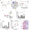
Figure 3. Parallel evolution of M. abscessus within and between patients.
(A) Example of within-host parallel evolution in P4 where several subclones have independently acquired a non-synonymous mutation in phoR (red), engA (yellow), and embC (pale blue). (B) Within-patient parallel evolution in 18 patients (each patient represented by a concentric circle) of 7 genes (chromosomal position relative to reference (ATCC19997) strain shown). Size of circles represent the number of non-synonymous SNPs (or any mutation in the case of 23S) present in each patient. (C) Manhattan plot identifying genes with more non-synonymous mutations than would be expected by chance across 201 patients (size of circle indicates the number of patients mutations were identified in used a one tailed binomial test. The p-values were corrected for multiple testing using Benjamini Hochberg method, with nonsignificant values (>0.01) shown in the shaded grey area. (D) Network analysis (using String) suggests that many of the genes undergoing parallel evolution may be functionally related. Edge thickness represents strength of evidence for direct interaction. (E) Impact of inducible CRISPRi knockdown of selected genes on M. abscessus survival in primary human macrophages at 2h (grey) and 24h (black) post infection (24h/2h ratios shown above), bacterial viability assessed by colony forming units (CFU). (F) Intracellular survival within primary human macrophages (at 2h (grey) and 24h (black) of wild type (WT) M. abscessus, PhoPR knockout (PhoPRD) mutants alone, or expressing empty vector (EV), wild type PhoPR (PhoPRwt) or PhoPR containing a patient-derived PhoR mutation (PhoPRmut). Experiments were performed in at least triplicate on at least three separate occasions. Results from representative experiments shown as mean ± s.e with statistical significance determined using two-tailed Student t-test (* p < 0.05, ** p <0.01. *** p < 0.001). (G,H) Infection of bENaC-tg mice with wild type (WT, black), PhoPRΔ (white), PhoPRΔ::PhoPRwt (blue) or PhoPRD::PhoPRmut (red) M. abscessus showing (G) bacterial burden in the lungs and (H) representative histology (arrows denote granuloma (left) and mycobacteria (right). Statistical significance determined using two-tailed Student t-test (* p < 0.05, ** p <0.01. *** p < 0.001).
Figure 4
Figure 4. Constrained evolution of M. abscessus.
(A) Transmission rates of mutations are compared between all non-unique SNPs (grey), non-synonymous non-sensor loop mutations in PhoR (pink), mutations conferring aminoglycoside or macrolide resistance in 16S and 23S rRNA respectively (green), non-synonymous PhoR sensor loop mutations (red), and non-synonymous mutations affecting GPL production (blue). Corresponding sizes clades with shared mutations (represented as number of patients per outbreak cluster) are also shown (right). (B, C) Phylogenetic tree of isolates from patients within transmission chains, showing examples of (B) transmission of adaptive sensor-loop SNPs and (C) preferential cross-infection by subclones with un-evolved phoR. (D) Impaired survival on fomites of (i) phoR knockout mutants (red) and (ii) rough isolates with GPL mutations (blue) compared to isogenic controls (black). Experiments were performed in at least triplicate on at least three separate occasions. Results from representative experiments shown as mean ± s.e with statistical significance determined using two-tailed Student t-test (* p < 0.05, ** p <0.01. *** p < 0.001).
Figure 5
Figure 5. Method for inferring subclone population structure.
(A) Bacterial colony sweeps were taken from culture of longitudinal samples for each patient and DNA extracted. (B) DNA was deep sequenced and minority variants called (see Methods for details). (C, D) We tracked allele frequencies over time and noticed that many variants followed the same frequency trajectory, suggesting that they form a haplotype or subclone. (E) We then used MV-trees to infer the underlying subclone population structure and subsequently pruned the resultant trees, given their acyclic directed graph relationship, using transitive reduction (see Methods for details).

Comment in

References

    1. Griffith DE, et al. An official ATS/IDSA statement: diagnosis, treatment, and prevention of nontuberculous mycobacterial diseases. Am J Respir Crit Care Med. 2007;175:367–416. - PubMed
    1. Floto RA, et al. Cystic Fibrosis Foundation and European Cystic Fibrosis Society Consensus Recommendations for the Management of Nontuberculous Mycobacteria in Individuals with Cystic Fibrosis. Thorax. 2016;71(Suppl 1):i1–i22. (2015) - PMC - PubMed
    1. Richards CJ, Olivier KN. Nontuberculous Mycobacteria in Cystic Fibrosis. Semin Respir Crit Care Med. 2019;40:737–750. - PubMed
    1. Martiniano SL, Nick JA, Daley CL. Nontuberculous Mycobacterial Infections in Cystic Fibrosis. Thorac Surg Clin. 2019;29:95–108. - PubMed
    1. Haworth CS, et al. British Thoracic society guidelines for the management of non-tuberculous mycobacterial pulmonary disease (NTM-PD) Thorax. 2017;72:ii1–ii64. - PubMed

Publication types

MeSH terms