Review
. 2021 Apr 29;30(160):200152.
doi: 10.1183/16000617.0152-2020. Print 2021 Jun 30.

Pulmonary granulomatosis of genetic origin

Affiliations
Review

Pulmonary granulomatosis of genetic origin

Sebastian F N Bode et al. Eur Respir Rev. .

Abstract

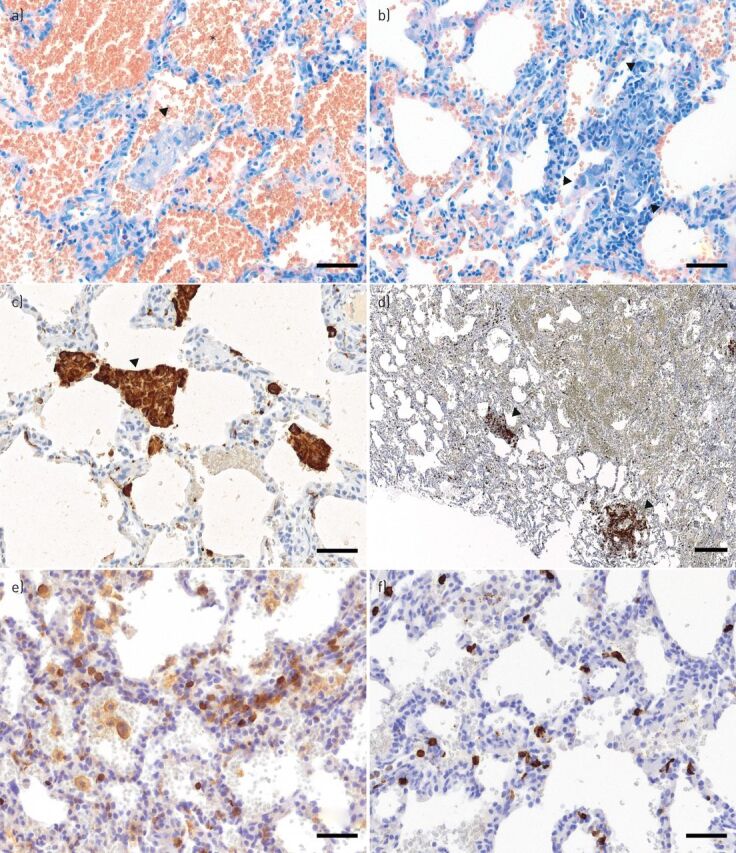
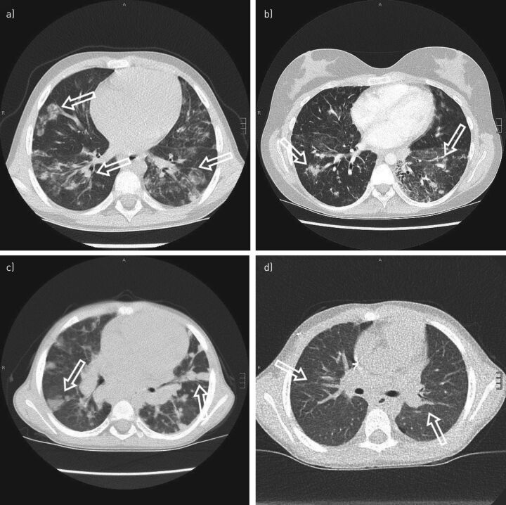
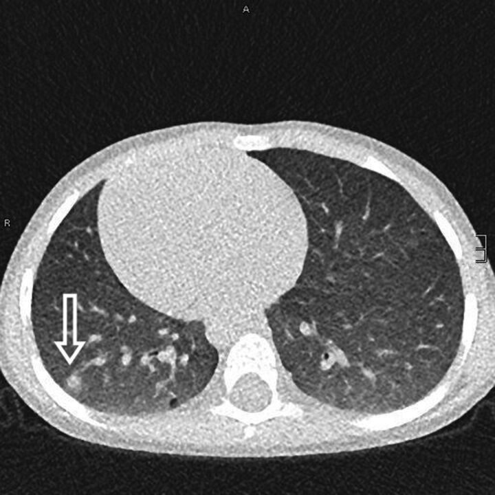
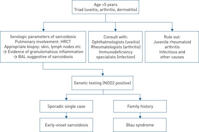
Granulomatous inflammation of the lung can be a manifestation of different conditions and can be caused by endogenous inflammation or external triggers. A multitude of different genetic mutations can either predispose patients to infections with granuloma-forming pathogens or cause autoinflammatory disorders, both leading to the phenotype of pulmonary granulomatosis. Based on a detailed patient history, physical examination and a diagnostic approach including laboratory workup, pulmonary function tests (PFTs), computed tomography (CT) scans, bronchoscopy with bronchoalveolar lavage (BAL), lung biopsies and specialised microbiological and immunological diagnostics, a correct diagnosis of an underlying cause of pulmonary granulomatosis of genetic origin can be made and appropriate therapy can be initiated. Depending on the underlying disorder, treatment approaches can include antimicrobial therapy, immunosuppression and even haematopoietic stem cell transplantation (HSCT). Patients with immunodeficiencies and autoinflammatory conditions are at the highest risk of developing pulmonary granulomatosis of genetic origin. Here we provide a review on these disorders and discuss pathogenesis, clinical presentation, diagnostic approach and treatment.


Conflict of interest statement

Conflict of interest: S.F.N. Bode has nothing to disclose. Conflict of interest: J. Rohr has nothing to disclose. Conflict of interest: J.M. Müller-Quernheim reports grants and non-financial support from Bristol Myers Squibb; personal fees from Roche and Novartis; personal fees and non-financial support from Boehringer Ingelheim; and non-financial support from CLS Behring, outside the submitted work. In addition, J.M. Müller-Quernheim has a patent “Use of inhaled VIP for treatment of immune checkpoint inhibitor-induced pneumonitis” pending and is co-founder of AdVita Lifescience GmbH, a spin-off of the University of Freiburg. Advita reports a pending intellectual property concerning the use of Aviptadil for the treatment of immune checkpoint inhibitor (ICI) pneumonitis. Conflict of interest: M. Seidl has nothing to disclose. Conflict of interest: C. Speckmann has nothing to disclose. Conflict of interest: A. Heinzmann has nothing to disclose.

Figures

FIGURE 1
FIGURE 1
Alveolar haemorrhage: partly epithelioid cellular interstitial inflammation in the affected lung, suggestive of a mild granulomatous-lymphocytic interstitial lung disease (GLILD). Airspaces are filled with erythrocytes (a), as indicated by an exemplar asterisk), some cluster forming alveolar macrophages (a) and c), as indicated by arrowheads, the latter using CD68+ stain) and small non-necrotising interstitial granulomas (b), as indicated by arrowheads) consisting of a few epithelioid cells and lymphocytes. Interstitial lymphocytic infiltrate mainly consists of CD4+ T-cells, partly clustered d) and partly scattered e). CD8+ T-cells are scattered f) and form the minority of the lymphocytic infiltrations. Scale bars: a–c, e, f) 50 μm; d) 200 μm.
FIGURE 2
FIGURE 2
Granulomatous-lymphocytic interstitial lung disease (GLILD). a) A 9-year old girl with atypical and confluent, partly spot-shaped infiltrates on chest computed tomography (CT) scan. b) A 20-year old woman showing nodules with adjacent ground-glass infiltrates. c) A chest CT scan of a 6-year old boy with cytotoxic T-lymphocyte-associated protein 4 deficiency. Multiple round infiltrates resembling “reversed halo” (or “atoll”) signs are evidence of GLILD. d) The same patient as in c) but 6 months after allogenic haematopoietic stem cell transplantation. Marked regression of GLILD is shown with mild residual, streaky interstitial consolidations. Scale bars: a, c, d) 15 mm; b) 20 mm.
FIGURE 3
FIGURE 3
Pulmonary granulomas in a 4-year old boy with chronic granulomatous disease. A subpleural granuloma with a surrounding fuzzy rim is indicated (arrow). Scale bar: 10 mm.
FIGURE 4
FIGURE 4
Proposed algorithm to differentiate between early onset sarcoidosis and Blau syndrome. HRCT: high-resolution computed tomography; BAL: bronchoalveolar lavage; NOD2: nucleotide-binding oligomerisation domain-containing protein 2. Data from [103].

References

    1. Ohshimo S, Guzman J, Costabel U, et al. . Differential diagnosis of granulomatous lung disease: clues and pitfalls. Eur Respir Rev 2017; 26: 170012. - PMC - PubMed
    1. Mukhopadhyay S, Gal AA. Granulomatous lung disease: an approach to the differential diagnosis. Arch Pathol Lab Med 2010; 134: 667–690. - PubMed
    1. Greenberg DE, Goldberg JB, Stock F, et al. . Recurrent Burkholderia infection in patients with chronic granulomatous disease: 11-year experience at a large referral center. Clin Infect Dis 2009; 48: 1577–1579. - PMC - PubMed
    1. Lieberman S, Gleason JB, Ilyas MIM, et al. . Assessing the safety and clinical impact of thoracoscopic lung biopsy in patients with interstitial lung disease. J Clin Diagn Res 2017; 11: OC57–OC59. - PMC - PubMed
    1. Blackhall V, Asif M, Renieri A, et al. . The role of surgical lung biopsy in the management of interstitial lung disease: experience from a single institution in the UK. Interact Cardiovasc Thorac Surg 2013; 17: 253–257. - PMC - PubMed

LinkOut - more resources