. 2021 Dec;76(12):1242-1245.
doi: 10.1136/thoraxjnl-2021-217031. Epub 2021 Apr 29.

Pulmonary fibrosis 4 months after COVID-19 is associated with severity of illness and blood leucocyte telomere length

Affiliations

Pulmonary fibrosis 4 months after COVID-19 is associated with severity of illness and blood leucocyte telomere length

Claire F McGroder et al. Thorax. 2021 Dec.

Abstract

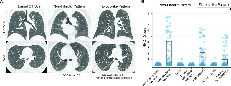
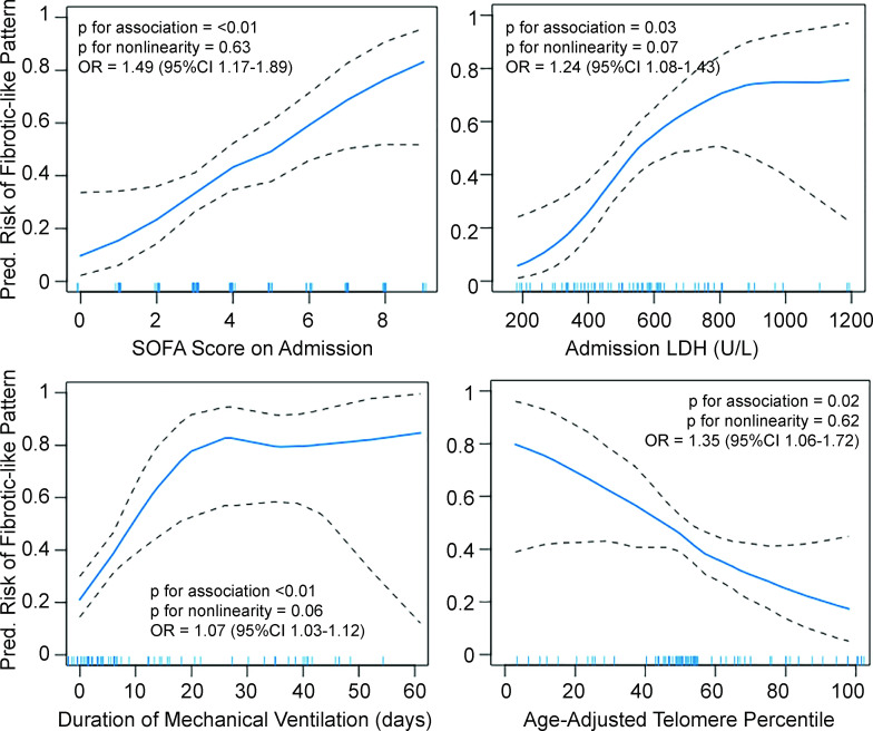
The risk factors for development of fibrotic-like radiographic abnormalities after severe COVID-19 are incompletely described and the extent to which CT findings correlate with symptoms and physical function after hospitalisation remains unclear. At 4 months after hospitalisation, fibrotic-like patterns were more common in those who underwent mechanical ventilation (72%) than in those who did not (20%). We demonstrate that severity of initial illness, duration of mechanical ventilation, lactate dehydrogenase on admission and leucocyte telomere length are independent risk factors for fibrotic-like radiographic abnormalities. These fibrotic-like changes correlate with lung function, cough and measures of frailty, but not with dyspnoea.

Keywords: COVID-19; imaging/CT MRI etc; interstitial fibrosis; respiratory measurement; viral infection.


Conflict of interest statement

Competing interests: None declared.

Figures

Figure 1
Figure 1
High-resolution CT (HRCT) scans of the chest from COVID-19 survivors. (A) Representative CT chest scans demonstrating no abnormalities (left), non-fibrotic patterns (middle) and fibrotic-like patterns (right). The upper panels show a coronal section and the lower panels show an axial image at the level just below the carina. The scan with a non-fibrotic pattern had a ground glass opacities (GGO) score of 5.6 (84th percentile in the group). The CT scan with a fibrotic-like pattern had a reticulation score of 6.4 (98th percentile), a traction bronchiectasis score of 5.0 (95th percentile) and no honeycombing. (B) Chest HRCT scores for radiographic patterns observed in the study cohort. The middle line of the boxplot represents the median score; bottom and top lines represent the 25th and 75th percentile, respectively. Where no lines are seen, the 25th, 50th and 75th percentile scores were all 0. The extent of each pattern was graded using a scoring system developed by ARDSnet. The possible range of scores was 0–20 from all categories of abnormalities, except traction bronchiectasis, which had a possible range of 0–5.
Figure 2
Figure 2
Continuous associations of fibrotic-like patterns with admission Sequential Organ Failure Assessment (SOFA) score (top left), lactate dehydrogenase (LDH) levels (top right), duration of mechanical ventilation in days (bottom left) and age-adjusted leucocyte telomere length percentile (bottom right) using generalised additive models with locally weighted smoothing (LOESS). Blue line represents predicted values. Black dashed lines are 95% CIs. Hash marks along the x-axis indicate individual study participants. Since there was no evidence for non-linearity in the generalised additive models, we estimated adjusted ORs using logistic regression models. All models are adjusted for a common set of potential confounders (age, sex, race/ethnicity, days since infection, body mass index, pack-years of smoking, treatment with steroids while hospitalised) and the other independent variables of interest (SOFA score, LDH, days of mechanical ventilation, telomere length). OR for SOFA score is per point increase, for LDH is per 50-point increase, for mechanical ventilation is per day increase, and for telomere length is per 10% decrease in percent-predicted value.

References

    1. Zhao Y-M, Shang Y-M, Song W-B, et al. . Follow-Up study of the pulmonary function and related physiological characteristics of COVID-19 survivors three months after recovery. EClinicalMedicine 2020;25:100463. 10.1016/j.eclinm.2020.100463 - DOI - PMC - PubMed
    1. Shah AS, Wong AW, Hague CJ, et al. . A prospective study of 12-week respiratory outcomes in COVID-19-related hospitalisations. Thorax 2021;76:402–4. 10.1136/thoraxjnl-2020-216308 - DOI - PubMed
    1. Hui DS, Wong KT, Ko FW, et al. . The 1-year impact of severe acute respiratory syndrome on pulmonary function, exercise capacity, and quality of life in a cohort of survivors. Chest 2005;128:2247–61. 10.1378/chest.128.4.2247 - DOI - PMC - PubMed
    1. Wong K-tak, Antonio GE, Hui DSC, et al. . Severe acute respiratory syndrome: thin-section computed tomography features, temporal changes, and clinicoradiologic correlation during the convalescent period. J Comput Assist Tomogr 2004;28:790–5. 10.1097/00004728-200411000-00010 - DOI - PubMed
    1. Huang Y, Tan C, Wu J, et al. . Impact of coronavirus disease 2019 on pulmonary function in early convalescence phase. Respir Res 2020;21:163. 10.1186/s12931-020-01429-6 - DOI - PMC - PubMed

Publication types