Review
. 2021 Apr 30;6(1):170.
doi: 10.1038/s41392-021-00554-y.

Cytosolic DNA sensing by cGAS: regulation, function, and human diseases

Affiliations
Review

Cytosolic DNA sensing by cGAS: regulation, function, and human diseases

Le Yu et al. Signal Transduct Target Ther. .

Abstract

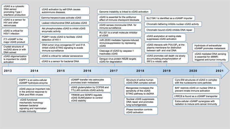
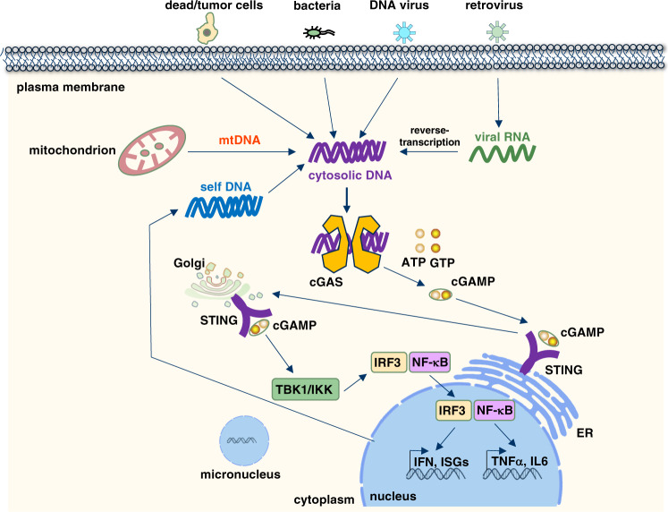
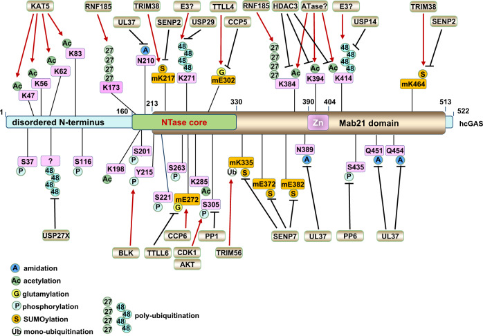
Sensing invasive cytosolic DNA is an integral component of innate immunity. cGAS was identified in 2013 as the major cytosolic DNA sensor that binds dsDNA to catalyze the synthesis of a special asymmetric cyclic-dinucleotide, 2'3'-cGAMP, as the secondary messenger to bind and activate STING for subsequent production of type I interferons and other immune-modulatory genes. Hyperactivation of cGAS signaling contributes to autoimmune diseases but serves as an adjuvant for anticancer immune therapy. On the other hand, inactivation of cGAS signaling causes deficiency to sense and clear the viral and bacterial infection and creates a tumor-prone immune microenvironment to facilitate tumor evasion of immune surveillance. Thus, cGAS activation is tightly controlled. In this review, we summarize up-to-date multilayers of regulatory mechanisms governing cGAS activation, including cGAS pre- and post-translational regulations, cGAS-binding proteins, and additional cGAS regulators such as ions and small molecules. We will also reveal the pathophysiological function of cGAS and its product cGAMP in human diseases. We hope to provide an up-to-date review for recent research advances of cGAS biology and cGAS-targeted therapies for human diseases.


Conflict of interest statement

The authors declare no competing interests.

Figures

Fig. 1
Fig. 1. A timeline for discoveries of cGAS regulation and function.
Due to a large amount of work on this topic in the past 8 years, we cannot include all major findings in this time table and we sincerely apologize for colleagues whose important work are not mentioned in this figure due to space constraints
Fig. 2
Fig. 2. The cGAS–STING signaling pathway senses cytosolic DNA derived from either viral/bacterial infection or self-DNA.
DNA is a pathogen-associated molecular pattern when delivered to the host cytoplasm by viral or microbial infection, and a danger-associated molecular pattern when leaked into the cytoplasm from damaged mitochondria or nucleus. cGAS is the cytosolic DNA sensor that recognizes and binds cytosolic DNA in a DNA-sequence-independent manner that subsequently triggers cGAS dimerization and production of a special dinucleotide messenger, 2′3′-cGAMP from ATP and GTP. 2′3′-cGAMP binds STING localized on ER, through trafficking to Golgi to recruit and activate IKK and TBK1. TBK1 phosphorylates STING, which in turn recruits IRF3 for phosphorylation by TBK1. Phosphorylated IRF3 dimerizes and enters the nucleus, where it cooperates with NF-κB signaling to turn on transcription of type I IFNs and other immunomodulatory genes
Fig. 3
Fig. 3. Regulations of cGAS activation by cGAS post-translational modifications.
cGAS is dynamically regulated by various post-translational modifications in responding to DNA insults, such as monoubiquitination, polyubiquitination, SUMOylation, glutamylation, phosphorylation, acetylation, and deamidation in cells. This figure illustrates up-to-date reported post-translational modifications occurring on cGAS proteins, including the modified residues and modifying enzymes. Please note that all human cGAS residues are labeled in pink while all mouse cGAS residues are labeled in orange with “m” inserted in front of the residue

References

    1. Hu MM, Shu HB. Cytoplasmic mechanisms of recognition and defense of microbial nucleic acids. Annu. Rev. Cell Dev. Biol. 2018;34:357–379. doi: 10.1146/annurev-cellbio-100617-062903. - DOI - PubMed
    1. Akira S, Uematsu S, Takeuchi O. Pathogen recognition and innate immunity. Cell. 2006;124:783–801. doi: 10.1016/j.cell.2006.02.015. - DOI - PubMed
    1. Atianand MK, Fitzgerald KA. Molecular basis of DNA recognition in the immune system. J. Immunol. 2013;190:1911–1918. doi: 10.4049/jimmunol.1203162. - DOI - PMC - PubMed
    1. Holm CK, Paludan SR, Fitzgerald KA. DNA recognition in immunity and disease. Curr. Opin. Immunol. 2013;25:13–18. doi: 10.1016/j.coi.2012.12.006. - DOI - PMC - PubMed
    1. Kagan JC. Signaling organelles of the innate immune system. Cell. 2012;151:1168–1178. doi: 10.1016/j.cell.2012.11.011. - DOI - PMC - PubMed

Publication types

MeSH terms