. 2021 Apr 29;12(1):2465.
doi: 10.1038/s41467-021-22769-x.

Scalable photonic-based nulling interferometry with the dispersed multi-baseline GLINT instrument

Affiliations

Scalable photonic-based nulling interferometry with the dispersed multi-baseline GLINT instrument

Marc-Antoine Martinod et al. Nat Commun. .

Abstract

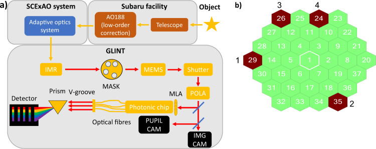
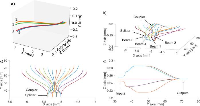
Characterisation of exoplanets is key to understanding their formation, composition and potential for life. Nulling interferometry, combined with extreme adaptive optics, is among the most promising techniques to advance this goal. We present an integrated-optic nuller whose design is directly scalable to future science-ready interferometric nullers: the Guided-Light Interferometric Nulling Technology, deployed at the Subaru Telescope. It combines four beams and delivers spatial and spectral information. We demonstrate the capability of the instrument, achieving a null depth better than 10-3 with a precision of 10-4 for all baselines, in laboratory conditions with simulated seeing applied. On sky, the instrument delivered angular diameter measurements of stars that were 2.5 times smaller than the diffraction limit of the telescope. These successes pave the way for future design enhancements: scaling to more baselines, improved photonic component and handling low-order atmospheric aberration within the instrument, all of which will contribute to enhance sensitivity and precision.


Conflict of interest statement

The authors declare no competing interests.

Figures

Fig. 1
Fig. 1. Global schematic of GLINT and configuration of the apertures of the mask.
a Global schematic of GLINT and its integration within the Subaru Telescope and SCExAO. Within GLINT itself the sequence of optical systems encountered consists of the image rotator (IMR), a pupil mask, a steerable segmented mirror (MEMS), a shutter and a linear polarising beamsplitter (POLA). The science beam is then injected by a microlens array (MLA) into the photonic chip while the dichroic beamsplitter delivers light to cameras viewing both the image plane and pupil plane. The processed beams coming from the photonic chip are spectrally dispersed in the GLINT spectrograph then imaged on the science detector (C-Red 2). b Diagram of the segmented MEMS mirror with highlighted segments in red matching the pattern of holes in the mask. Segments 29, 35, 26 and 24 are identified with beams 1, 2, 3 and 4, respectively. The lengths of baselines range from 2.15 m (combination of the beams 3 and 4) to 6.45 m (combination of the beams 2 and 3).
Fig. 2
Fig. 2. Schematics of the integrated-optics chip.
a Schematics of the pupil remapper of the chip, coherently transforming the 2D configuration of the inputs (on the left) matching the desired pupil sampling pattern into a 1D configuration (on the right). The waveguide paths have been optimised to match their optical path lengths despite their different routes. The green waveguide is associated with beam 1, orange with beam 2, red with beam 3 and blue with beam 4. b Perspective view of the beam combiner of the chip. c Plan view in which light propagates from the 4 inputs at the bottom towards the top, encountering 4-way splitters and codirectional couplers. d Right-side view of the chip showing the locations of the inputs and the outputs. The inputs, outputs, splitters and couplers are indicated on the (bd) diagrams. The axis scale proportions in all the schematics differ for clarity in the drawing.
Fig. 3
Fig. 3. Scanned fringe envelopes for each of the six baselines.
Subplots (a to f) respectively represent the baselines N1 to N6 scanned over the full available range of the optical path difference (OPD) from data taken in July 2019 with illumination from the supercontinuum source within SCExAO. The blue dots are the data and the orange curve is the fitted model. The label of the horizontal axis “OPDxy” shows the segment of the beam x was scanned with respect to the reference beam y. The value in each plot is the fitted δ0.
Fig. 4
Fig. 4. Splitting, coupling ratios and ζ coefficients of the beam combiner.
a Splitting ratios of beam 1 between the photometric output and the three couplers with respect to wavelength. b Coupling ratios of the coupler combining the beams 1 and 2 with respect to wavelength. c ζ coefficients for beams 1 and 2 for the outputs of Null 1 with respect to wavelength.
Fig. 5
Fig. 5. A data frame from the science sensor illuminated with a laboratory source.
Spectral dispersion runs along the horizontal direction, while outputs from different waveguides are displaced in vertical rows. The rows labelled “P” are the photometric tap. “N1-6” are the outputs corresponding to the null output and “AN1-6” are the outputs corresponding to the respective anti-null outputs. On this occasion, pistons on the MEMS mirror were configured to produce a nulled signal on N1 and N4.
Fig. 6
Fig. 6. Source null depth with respect to baseline by observing the supercontinuum source of the SCExAO bench.
The blue dots represent the null depths measured from the binned spectral channel while the orange dots represent the null depths measured with spectrally dispersed light.
Fig. 7
Fig. 7. Histograms of the measured null depth of base N1 computed over 10 dispersed spectral channels with the fitted curves.
Each subplot from (a) to (j) shows the histogram of the measured null depth of base N1 on α Boo (blue dots) and the fitted model (orange curve), in each of the spectral channel spanning from 1525 to 1570 nm. The fitted parameters are Nobj,12 = 7.05 × 10−2 ± 1.86 × 10−4, μOPD = 302 ± 0.465 nm and σOPD = 163 ± 0.163 nm for a reduced χ2 of 2.51.
Fig. 8
Fig. 8. Variations of the source null depth of α Boo and δ Vir and their respective fitted curves with respect to the baseline.
a Variation of the source null depth of α Boo (blue dots) as a function of baseline together with the best-fit model (blue solid curve). The orange area highlights the expected range of null depth at 1550 nm for an expected UD diameter between 19.1 and 20.4 mas (obtained from the literature). Error bars represent the standard error of the measurements. b Source null depth of δ Vir (blue dots) with baseline together with the best-fit model (blue solid curve). The orange gives the expected range of null depth for a UD diameter between 9.86 and 11.3 mas (from the literature). The error bars represent the standard deviation of the fitted value of the source null depth given by the covariance matrix of the fit of the histogram, rescaled by the reduced χ2 of that fit.

References

    1. Schneider J, Dedieu C, LeSidaner P, Savalle R, Zolotukhin I. Defining and cataloging exoplanets: the exoplanet.eu database. A&A. 2011;532:A79. doi: 10.1051/0004-6361/201116713. - DOI
    1. Fujii Y, et al. Colors of a second earth: estimating the fractional areas of ocean, land, and vegetation of earth-like exoplanets. ApJ. 2010;715:866–880. doi: 10.1088/0004-637X/715/2/866. - DOI
    1. Kawahara H, et al. Can ground-based telescopes detect the oxygen 1.27 μm absorption feature as a biomarker in exoplanets? ApJ. 2012;758:13. doi: 10.1088/0004-637X/758/1/13. - DOI
    1. Seager S, Bains W, Petkowski JJ. Toward a list of molecules as potential biosignature gases for the search for life on exoplanets and applications to terrestrial biochemistry. Astrobiology. 2016;16:465–485. doi: 10.1089/ast.2015.1404. - DOI - PubMed
    1. Guyon, O. et al. How ELTs will acquire the first spectra of rocky habitable planets, volume 8447 of Society of Photo-Optical Instrumentation Engineers (SPIE) Conference Series, 84471X, 10.1117/12.927181 (2012).

LinkOut - more resources