. 2021 Apr 29;11(1):9301.
doi: 10.1038/s41598-021-88902-4.

Theileria equi claudin like apicomplexan microneme protein contains neutralization-sensitive epitopes and interacts with components of the equine erythrocyte membrane skeleton

Affiliations

Theileria equi claudin like apicomplexan microneme protein contains neutralization-sensitive epitopes and interacts with components of the equine erythrocyte membrane skeleton

Cynthia K Onzere et al. Sci Rep. .

Abstract

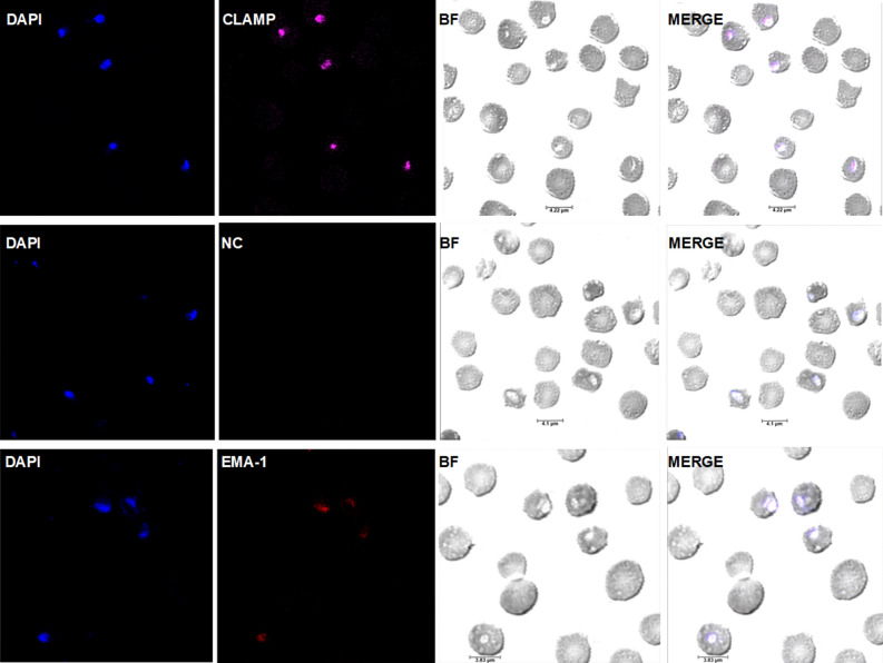
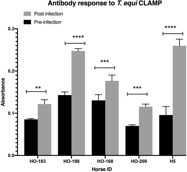
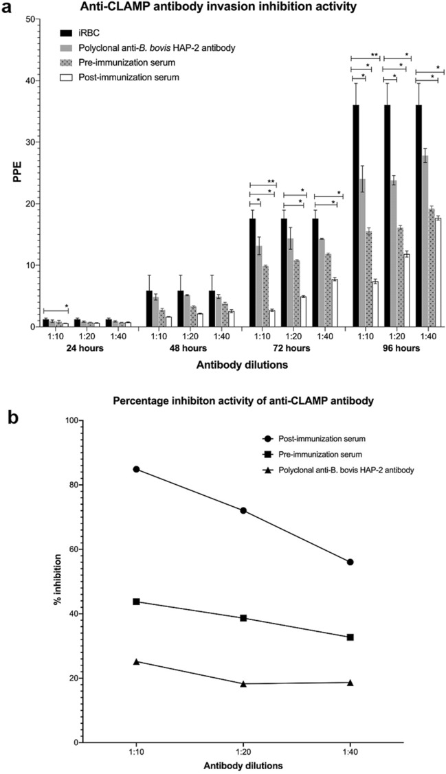
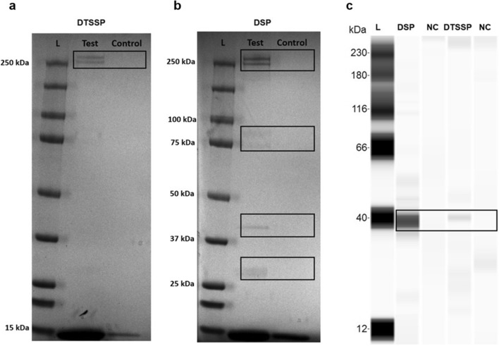
Theileria equi is a widely distributed apicomplexan parasite that causes severe hemolytic anemia in equid species. There is currently no effective vaccine for control of the parasite and understanding the mechanism that T. equi utilizes to invade host cells may be crucial for vaccine development. Unlike most apicomplexan species studied to date, the role of micronemes in T. equi invasion of host cells is unknown. We therefore assessed the role of the T. equi claudin-like apicomplexan microneme protein (CLAMP) in the invasion of equine erythrocytes as a first step towards understanding the role of this organelle in the parasite. Our findings show that CLAMP is expressed in the merozoite and intra-erythrocytic developmental stages of T. equi and in vitro neutralization experiments suggest that the protein is involved in erythrocyte invasion. Proteomic analyses indicate that CLAMP interacts with the equine erythrocyte α-and β- spectrin chains in the initial stages of T. equi invasion and maintains these interactions while also associating with the anion-exchange protein, tropomyosin 3, band 4.1 and cytoplasmic actin 1 after invasion. Additionally, serological analyses show that T. equi-infected horses mount robust antibody responses against CLAMP indicating that the protein is immunogenic and therefore represents a potential vaccine candidate.


Conflict of interest statement

The authors declare no competing interests.

Figures

Figure 1
Figure 1
T. equi CLAMP contains immunogenic epitopes, and the gene is transcribed and expressed by T. equi merozoites. (a) CLAMP’s transmembrane topology as predicted and annotated by Phobius and Protter algorithms, respectively. Peptides 1, 2 and 3 indicate the protein’s predicted immunogenic peptides. (b) A 1% agarose gel showing amplified clamp transcript at ~ 1226 bp. The T. equi equine merozoite antigen-1 (ema-1) transcript was amplified as a positive control, and rt- (negative control) represents amplified merozoites’ RNA without addition of reverse transcriptase. L indicates the Thermo Scientific GeneRuler 1 kb DNA Ladder. (c) Immunoblot showing CLAMP expressed by T. equi merozoites (test) at ~ 39 kDa. Probing of merozoites lysate with pre-immunization serum and the polyclonal anti-RAP-1a antibody were used as negative control (NC) and positive control (PC) respectively. L represents the ProteinSimple Wes Ladder.
Figure 2
Figure 2
CLAMP is expressed on the surface of T. equi merozoites. Confocal microscopy images showing expression of CLAMP by extracellular T. equi merozoites. The first row shows expression of CLAMP on the surface of an extracellular merozoite (indicated by an asterisk (*)). The nuclei are stained in blue with DAPI, and CLAMP is stained in magenta. The second row represents merozoites incubated with the pre-immunization serum (negative control (NC)) and shows lack of protein staining indicating that the staining in the first row was CLAMP-specific. The third row shows expression of EMA-1 (positive control (PC)) by merozoites. In all cases, BF refers to brightfield panels.
Figure 3
Figure 3
CLAMP is expressed in the intra-erythrocytic stages of T. equi development. Confocal microscopy images showing expression of CLAMP in T. equi infected erythrocytes. The first row shows expression of CLAMP by T. equi gamonts. Parasite nuclei are stained in blue with DAPI, and CLAMP is stained in magenta. The second row shows infected erythrocytes incubated with pre-immunization serum (negative control) and it shows lack of staining indicating test specificity. The third row indicates expression of EMA-1 (positive control (PC)) by intra-erythrocytic parasites. In all cases, BF refers to brightfield panels.
Figure 4
Figure 4
CLAMP elicits antibody responses in horses during infection with T. equi. CLAMP-specific antibodies are significantly present in horses after infection with T. equi (post- infection). **p = 0.0014, ***p = 0.0002, ****p < 0.0001.
Figure 5
Figure 5
Polyclonal anti-CLAMP antibody significantly inhibits invasion of equine erythrocytes by T. equi. (a) In vitro neutralization assay data showing invasion inhibition activity of the CLAMP-specific antibody (post-immunization serum), pre-immunization serum and polyclonal anti-B. bovis HAP-2 antibody at different dilutions over time. T. equi infected erythrocytes cultured in the presence of CLAMP-specific antibodies were significantly less parasitized (PPE) compared to parasites cultured in the presence of pre-immunization serum and the polyclonal anti-B. bovis HAP-2 antibody. Control represents T. equi infected red blood cells (iRBC) cultured in the absence of both pre- and post-immunization sera. *p < 0.05, **p < 0.0090. (b) Anti-CLAMP antibodies inhibit a significant percentage of parasites from invading erythrocytes at 72 h post-infection. Percentage inhibition was calculated as the difference between percentage inhibition in the presence of polyclonal anti-CLAMP antibodies (post-immunization serum) and percentage inhibition in the presence of pre-immunization serum. In this regard, anti-CLAMP antibodies in the post-immunization serum inhibited parasite invasion by 41%, 33.4% and 23.3% at 1:10, 1:20 and 1: 40 dilutions, respectively. Factors present in the B. bovis HAP-2 post immunization serum (polyclonal anti-B. bovis HAP-2 antibody) also minimally inhibited invasion.
Figure 6
Figure 6
T. equi CLAMP interacts with equine erythrocyte proteins. (a,b) show SDS-PAGE gels of proteins (within black boundaries) isolated from the erythrocyte surface (test) and within the cell membrane and cell (test) after crosslinking to CLAMP with DTSSP and DSP, respectively. (c) Represents an immunoblot showing the presence of CLAMP at ~ 39 kDa. In all cases agarose resin eluates were used as negative controls (control/NC). The ~ 15 kDa band on the SDS-PAGE gels indicate non-specific binding of the equine α- and β-hemoglobin subunits to the Thermo Scientific AminoLink (test) and agarose (control) coupling resins.
Figure 7
Figure 7
Hypothetical model showing the role of T. equi CLAMP in invasion of equine erythrocytes and its interactions with membrane skeleton proteins. (a) T. equi attaches to the equine erythrocyte surface, and (b) reorientates itself such that the apical end is in direct association with the cell membrane. (c) The parasite uses CLAMP (that is possibly translocated from the micronemes to the cell surface) to attach to the α- and β-spectrin proteins during the initial stages of invasion. (d) Once inside the cell, T. equi CLAMP maintains its interactions with the spectrin proteins while also interacting with band 4.1, tropomyosin 3, cytoplasmic actin 1 and anion-exchange protein (AE).

Similar articles

Cited by

References

    1. Wise LN, Kappmeyer LS, Mealey RH, Knowles DP. Review of equine piroplasmosis. J. Vet. Intern. Med. 2013;27:1334–1346. doi: 10.1111/jvim.12168. - DOI - PubMed
    1. Ueti MW, Palmer GH, Scoles GA, Kappmeyer LS, Knowles DP. Persistently infected horses are reservoirs for intrastadial tick-borne transmission of the apicomplexan parasite Babesia equi. Infect. Immun. 2008;76:3525–3529. doi: 10.1128/IAI.00251-08. - DOI - PMC - PubMed
    1. Scoles GA, et al. Piroplasmosis associated with amblyomma. Emerg. Infect. Dis. 2011;17:1903–1905. doi: 10.3201/eid1710.101182. - DOI - PMC - PubMed
    1. Bargieri D, Lagal V, Andenmatten N, Tardieux I, Meissner M. Host cell invasion by apicomplexan parasites: The junction conundrum. PLoS Pathog. 2014;10:1–9. doi: 10.1371/journal.ppat.1004273. - DOI - PMC - PubMed
    1. Carruthers V. B., S. L. D. Sequential protein secretion from three distinct organelles of Toxoplasma gondii accompanies invasion of human fibroblasts. Eur. J. Cell Biol.73, 114–123 (1997). - PubMed

Publication types

MeSH terms

LinkOut - more resources