

doi: 10.1038/s41594-021-00585-7.
Epub 2021 Apr 29.
The structure of a virus-encoded nucleosome
Affiliations
- PMID: 33927388
- PMCID: PMC8370576
- DOI: 10.1038/s41594-021-00585-7
Item in Clipboard
The structure of a virus-encoded nucleosome
Nat Struct Mol Biol.
2021 May.
Abstract
Certain large DNA viruses, including those in the Marseilleviridae family, encode histones. Here we show that fused histone pairs Hβ-Hα and Hδ-Hγ from Marseillevirus are structurally analogous to the eukaryotic histone pairs H2B-H2A and H4-H3. These viral histones form 'forced' heterodimers, and a heterotetramer of four such heterodimers assembles DNA to form structures virtually identical to canonical eukaryotic nucleosomes.
Figures
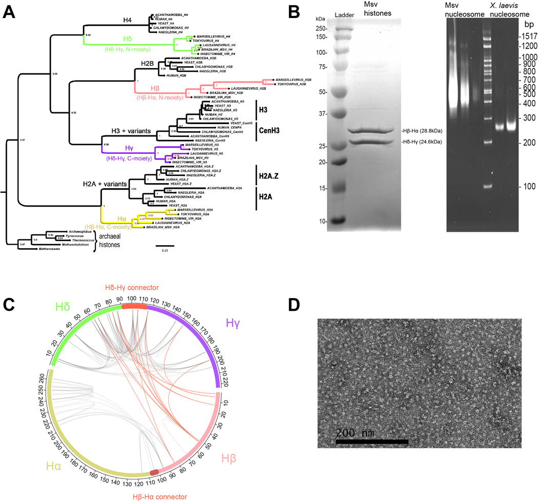
a, A protein sequence alignment based on the Marseillevirus histone heterotetrameric structure was used to infer the phylogenetic relationships of the separate histone moieties (N- and C- terminal halves) from each the two histone doublet genes from three different Marseilleviridae genomes. This tree shows that each of the four viral histone domains (Hβ and Hα from the Hβ-Hα gene, and Hδ and Hγ from the Hδ-Hγ gene) is related to one of the four canonical eukaryotic core histone families. Furthermore, the C-terminal halves of each viral histone doublet are distantly related to eukaryotic core histone sub-families that come in distinctive functional variants (Hα → H2A + variants, and Hγ → H3 + variants). This phylogeny was computed using Bayesian inference on 193 alignment columns (Materials & Methods). Histone domains encoded by various archaeal genomes were used as an outgroup clade to determine root placement. b, SDS-PAGE gel of Msv histones stained with Coomassie blue (left) and PAGE native gel with different dilutions of Msv and Xenopus nucleosome assemblies stained with ethidium bromide (right). Each sample was run independently twice (n=2). c, Crosslinking mass spectrometry revealed intramolecular (gray lines) and intermolecular (red lines) contacts between histones. d, Negative stain electron microscopy image of glutaraldehyde crosslinked viral histone assemblies. This image shows presence of round, nucleosome shaped particles.
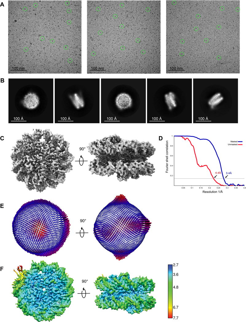
Data were collected on Titan Krios 300kV microscope. a, Raw cryo-EM images of Msv nucleosome were collected as described in Materials and Methods. Individual particles are highlighted in green circles. b, Representative 2D class averages selected from the dataset. c, Two representative views of the 3.4 Å 3D reconstruction Msv nucleosome. d, Fourier Shell Correlation plot of the 3.4 Å Msv nucleosome between two independently refined half maps (measured at FSC=0.143). e, Euler angle distribution of assignment of particles used to generate the final 3.4 Å reconstruction. The length of every cylinder is proportional to the number of particles assigned to the specific orientation. f, Two different views of the heat map of the Msv nucleosome cryo-EM density colored by local resolution.
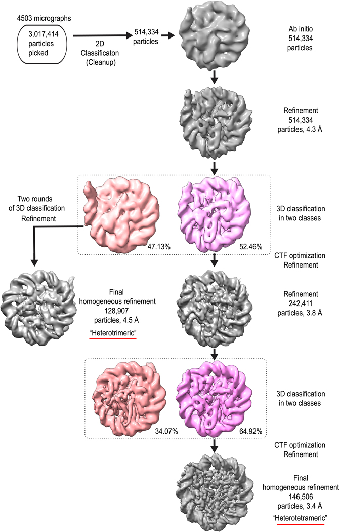
Detailed summary of classification of Msv dataset collected on Titan Krios operated at 300 kV. The two classes from the first 3D classification were further processed to leading to the “heterotetrameric” and “heterotrimeric” Msv nucleosomes.
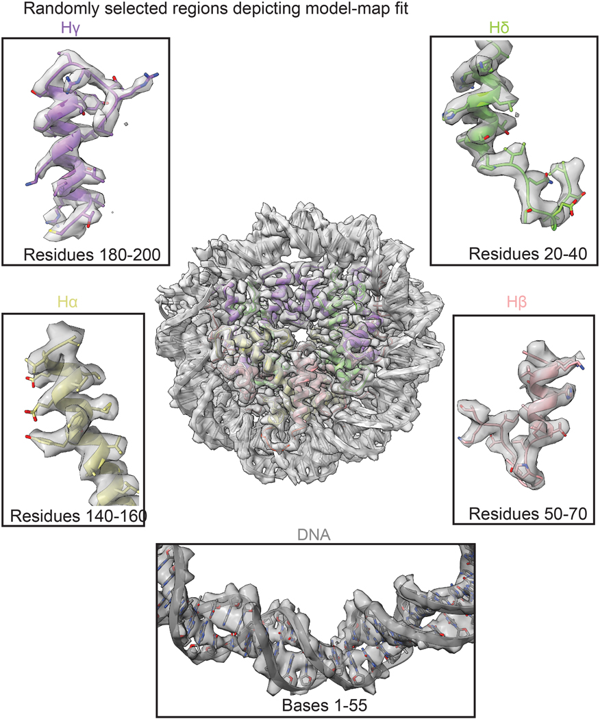
Fitting of the model to the Cryo-EM density of different regions of the Msv histones Hα, Hβ, Hγ, Hδ. The model is color coded as in Fig. 1.
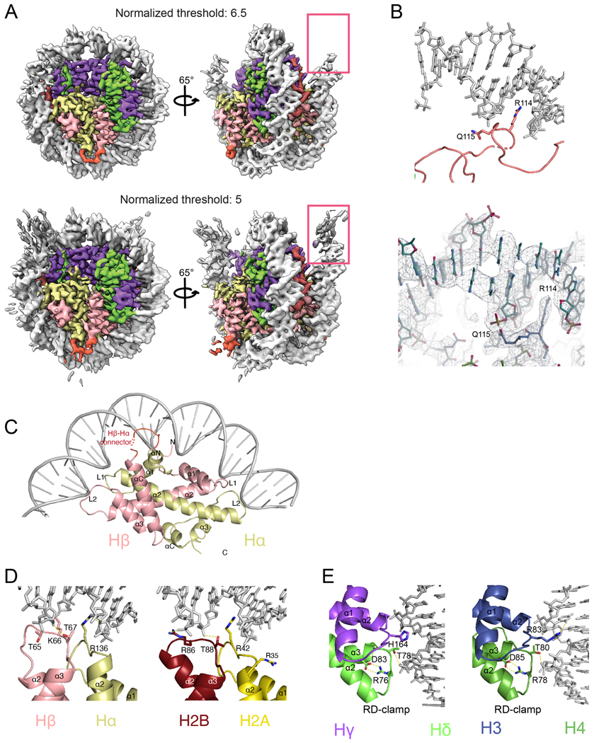
a, Cryo-EM density of Msv nucleosome at two different thresholds showing the flexibility of the DNA ends in the Msv nucleosome. Two different views of the Coulomb map at contouring level of 6.5 sigma (top) and 5 sigma (bottom). At a lower contouring, the full length of the DNA (147 bp) is still visible but only 121 bp are stabilized and coordinates can be assigned. The map is color coded as in Fig. 1. b, Close up of the model and cryo-EM map fitting of specific contacts between the Hδ-Hγ connector and the DNA. c, Overview of the Hβ-Hα DNA interface. d, Comparison of interactions between Hβ-Hα with DNA (left) and H2B and H2A with DNA (right). e, Interactions of Hδ-Hγ with the DNA (left) and H3 and H4 with the DNA (right). Human nucleosome (PDB ID 5AV9).
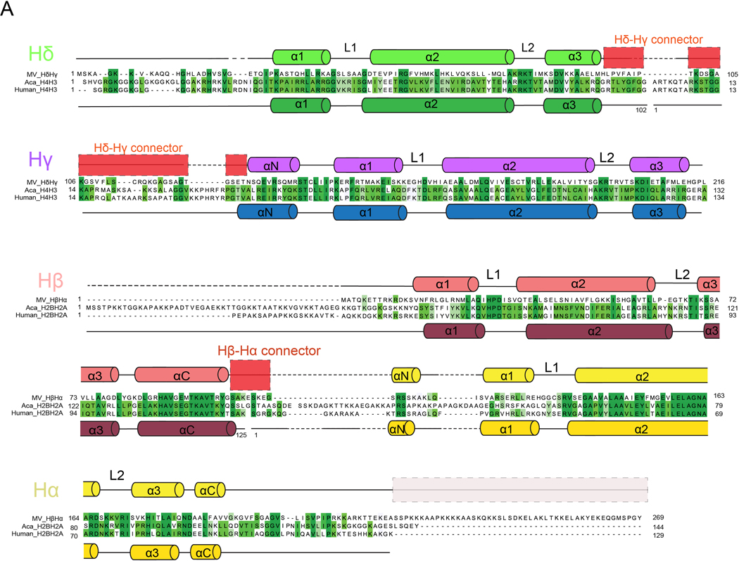
Sequence alignment depicting secondary structures of Msv, human and A. castellanii histones. The secondary structure is shown for Msv and Human histones only. No structural data is available for A. castellanii. Shades of green indicate identical (dark green) and similar (light green) residues. Sequence alignments were performed with Clustal Omega from UniPort. A summary of structural and sequence alignments is shown in Extended Data Fig. 10.
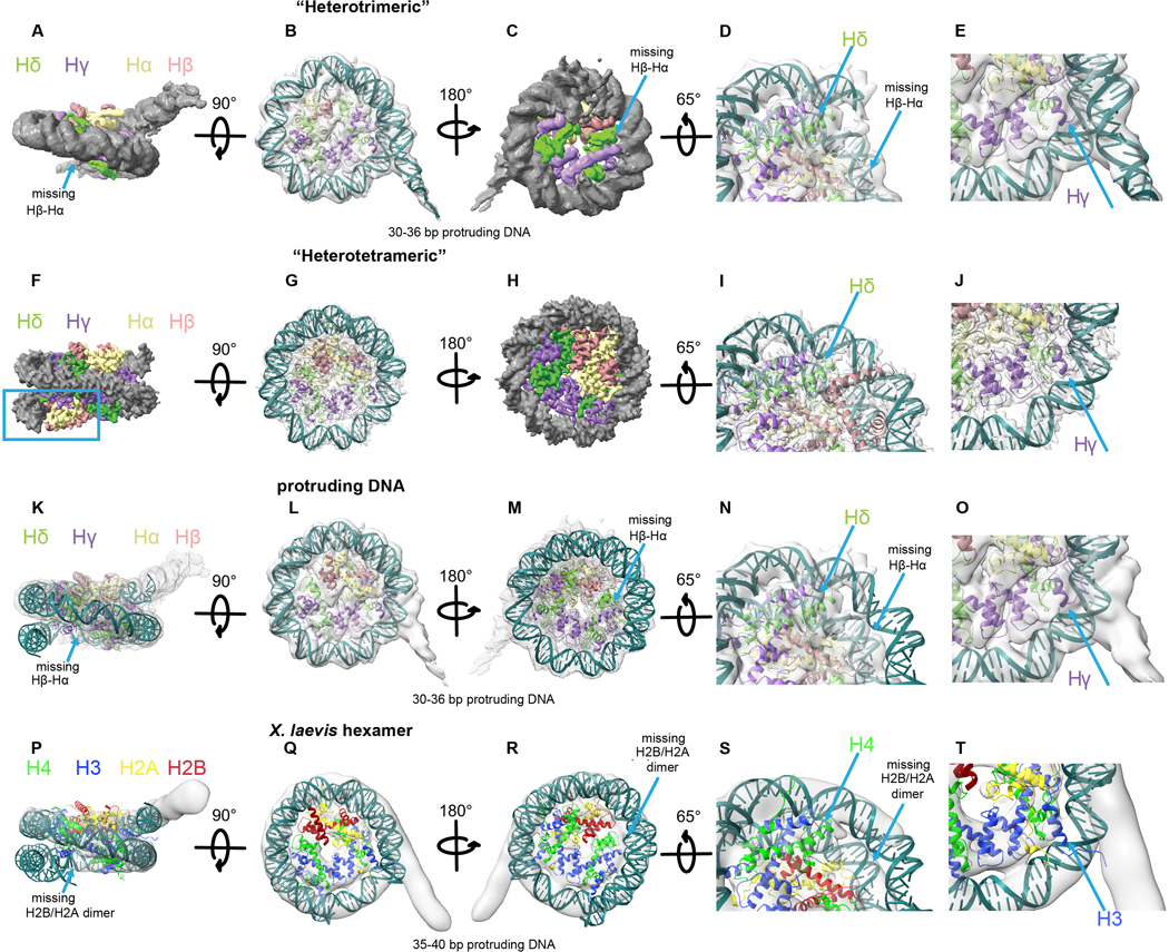
a-e, Model of the “heterotrimeric” Msv nucleosome with modeled DNA fitting inside the “heterotrimeric” Msv cryo-EM map. a-c, Three different views, d, close up of the region where DNA is missing and e, close-up of the region where DNA is protruding. f-j, Model of the “heterotetrameric” Msv nucleosome fitting inside the “heterotetrameric” Msv cryo-EM map. f-h, Three different views, i and j, close ups of the regions shown in d and e. k-o, Model of the “heterotrimeric” Msv nucleosome without the modeled DNA fitting inside the “heterotrimeric” Msv cryo-EM map. k-m, Three different views, n and o, close ups of the regions shown in d and e. p-t, Docking of a model of a “hexameric” eukaryotic nucleosome (PDB ID 5AV9) produced taking off one of the dimers H2B and H2A and fitted inside the “hexameric” Xenopus cryo-EM map EMD-3929. Please note that we did not rebuilt the DNA in this model. p-r, Three different views, s and t,. close ups of the regions shown in d and e. Maps and models are color coded as Fig. 1. The blue square shows the missing DNA region and the arrow the missing Hβ-Hα (or H2B and H2A) histones.
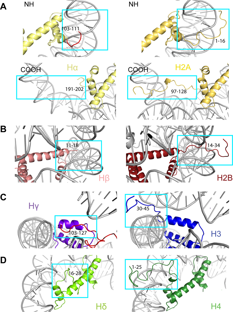
Comparison of the N-terminal tails of the Msv nucleosome with the Xenopus nucleosome (PDB ID 1KX5) for: a, N-terminal Hα (top left) vs H2A (top right), C-terminal Hα (bottom left) vs H2A (bottom right). b, N-terminal Hβ vs H2B, c, N-terminal Hγ vs H3 and d, N-terminal Hδ vs H4. The models are color coded as in Fig. 1. The histone tail residues are indicated.
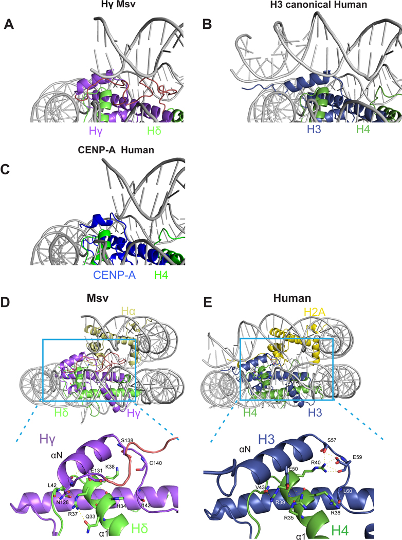
Comparison of interactions between DNA terminus with a, Hγ in Msv, b, canonical H3 in human (PDB ID 5AV9) and c, CENP-A in centromeric human nucleosomes (PDB ID 3AN2). d-e, Overview (top) and close-up (bottom) of the contacts established between Hδ α1 helix and Hγ αN helix of the d, Msv and e, human nucleosome (PDB ID 5AV9). Differences in the interface between Msv Hδ α1 helix with Hγ αN helix when compared to H4 α1 helix with H3 αN helix could contribute to increased flexibility of the DNA ends in the Msv nucleosome. The nucleosome is color coded as in Fig. 1.
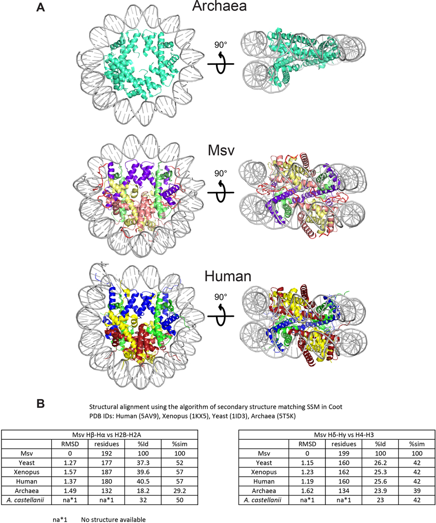
a, Two different views of (top) archaea (PDB ID 5T5K), (middle) Msv and (bottom) human (PDB ID 5AV9) nucleosomes. b, RMSD and sequence conservation between histones in different organisms. Structural alignment and identity were obtained using the SSM algorithm in Coot. All the values of similarity and the sequence alignment with A. castellanii were obtained with BLASTp from NCBI. Only one homodimer from Archaea was possible to be aligned to the heterodimers for other organisms.
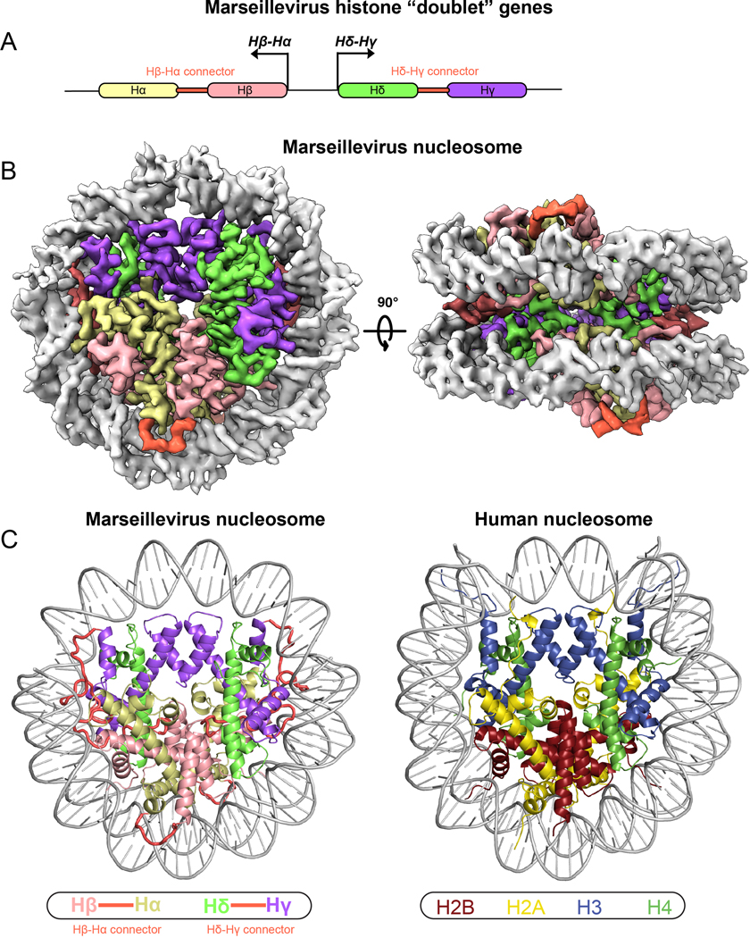
a, Cartoon depiction of divergently transcribed histone “doublet” genes Hβ-Hα and Hδ-Hγ. b, Cryo-EM reconstruction of viral “nucleosomes” displayed in two separate views related by 90°. c, Comparison of the Marseillevirus nucleosome cryo-EM structure (left) and the human nucleosome (right, PDB ID 5AV9).
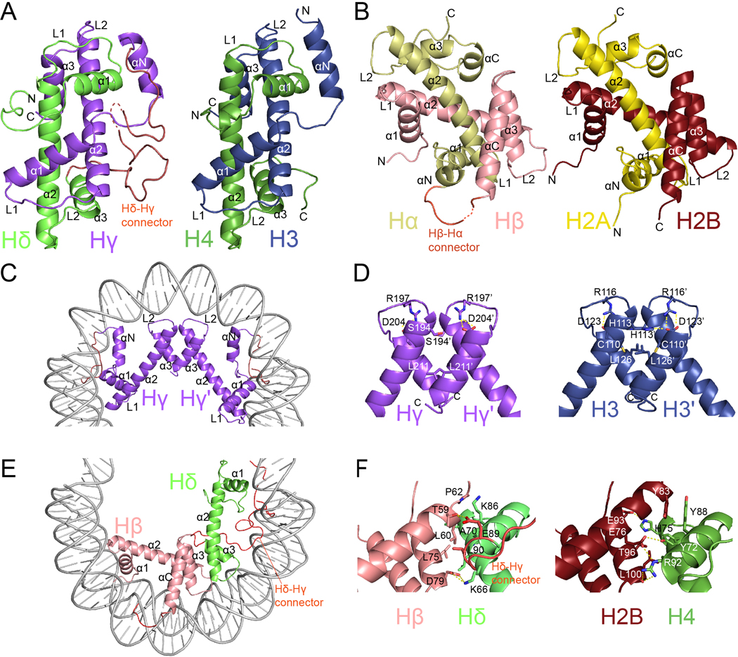
Comparison of Msv histone “forced dimers” (a and b
left) with human histone heterodimers (a and b
right). Overview of c, Hγ-Hγ’ and e, Hβ-Hδ four helix bundles. d, Detailed view of Hγ-Hγ’ (left) and H3-H3’ (right) interfaces f, Detailed view of Hβ-Hδ (left) and H2B-H4 (right) interfaces.

a Hδ-Hγ “forced dimer”-DNA interface. b, Comparison of interactions between Hδ-Hγ with DNA (left) and human H4 and H3 with DNA (right). c, Close-up view of αN helices and the DNA ends of the Hγ (left) and H3 (right). d, Overview of the C-termini of Hα (left) and H2A (right). e, Representation of electrostatic surface potential of Msv (left) and human (right) nucleosomes. f, Close-up of the acidic patch in Marseillevirus Hβ-Hα (left) and H2B and H2A (right).
Comment in
-
A small nucleosome from a weird virus with a fat genome.Mol Cell. 2021 Sep 2;81(17):3447-3448. doi: 10.1016/j.molcel.2021.08.014. Mol Cell. 2021. PMID: 34478653
References
-
- Talbert PB, Meers MP & Henikoff S. Old cogs, new tricks: the evolution of gene expression in a chromatin context. Nat Rev Genet 20, 283–297 (2019). - PubMed
-
- Kornberg RD Chromatin structure: a repeating unit of histones and DNA. Science 184, 868–71 (1974). - PubMed
-
- Luger K, Mader AW, Richmond RK, Sargent DF & Richmond TJ Crystal structure of the nucleosome core particle at 2.8 A resolution. Nature 389, 251–60 (1997). - PubMed
Publication types
MeSH terms
Substances
Grants and funding
LinkOut - more resources
Full Text Sources
Other Literature Sources
Molecular Biology Databases
