. 2021 Apr;12(2):402-409.
doi: 10.1055/s-0041-1727300. Epub 2021 Apr 23.

Magnetic Resonance Imaging in Peripartum Encephalopathy: A Pictorial Review

Affiliations

Magnetic Resonance Imaging in Peripartum Encephalopathy: A Pictorial Review

Duraipandi Manjubashini et al. J Neurosci Rural Pract. 2021 Apr.

Abstract

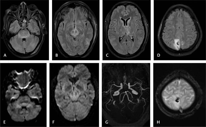
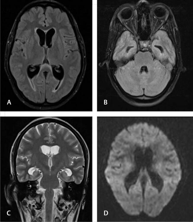
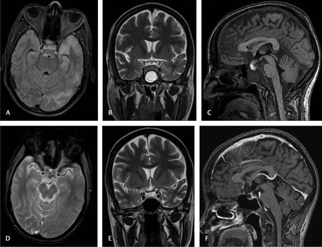
Acute neurological problems are significant disorders of pre- and postpartum period in women. We analyzed the magnetic resonance imaging (MRI) profile of patients presenting with peripartum encephalopathy over 2 years. Of 51 patients, MRI was abnormal in 40 patients and normal in 11 patients. Posterior reversible encephalopathy (13/40), cerebral venous thrombosis (6/40), and postpartum angiopathy (3/40) are the three most common causes of peripartum encephalopathy as identified in MRI. The other less common but important etiology include HELLP (Hemolysis, Elevated Liver enzymes and Low Platelet) syndrome (2), osmotic demyelination (2), antiphospholipid syndrome (2), tubercular meningitis/cerebritis (3), pituitary hyperplasia with hemorrhage (2), postictal edema (2), cerebellitis (1), transient splenial lesion (1), and changes of old trauma and stroke (one each).

Keywords: magnetic resonance imaging; peripartum encephalopathy; posterior reversible encephalopathy; postpartum angiopathy.


Conflict of interest statement

Conflict of Interest None declared.

Figures

Fig. 1
Fig. 1
Posterior reversible encephalopathy syndrome. A 21-year-old primigravida presented with postpartum encephalopathy. Fluid-attenuated inversion recovery axial sections ( A and B ) showing occipital and frontoparietal subcortical white matter hyperintensities. No diffusion restriction is noted ( C ). Magnetic resonance angiogram is normal ( D ).
Fig. 2
Fig. 2
Cerebral venous thrombosis. A 27-year-old female with second pregnancy presented before labor with altered sensorium. Fluid-attenuated inversion recovery axial ( A ), T2 coronal sections showing left temporoparietal hemorrhagic infarction due to venous thrombosis ( B ). Gradient-echo T2* ( C ) images showing hypointense hemorrhage with absent left transverse sinus in magnetic resonance venogram ( D ).
Fig. 3
Fig. 3
Postpartum angiopathy. A 23-year-old primigravida presented with encephalopathy. Fluid-attenuated inversion recovery axial, diffusion ( A , B ) and apparent diffusion coefficient ( C ) images shows acute infarction in the pons with areas if hemorrhage in gradient-echo T2* image ( D ). Magnetic resonance angiogram ( E ) is normal. Follow-up magnetic resonance imaging ( F ) after 6 weeks shows partial resolution.
Fig. 4
Fig. 4
Hemolysis, elevated liver enzymes, and low platelet. A 25-year-old primigravida presented 2 days before labor with encephalopathy, elevated liver enzymes, and mild reduction in platelet count. Fluid-attenuated inversion recovery axial sections ( AD ) showing hyperintensities in pons, midbrain, left caudate, thalamus, and right parietal regions. Subtle diffusion restriction noted in pons and midbrain lesions ( E and F ). Magnetic resonance angiogram ( G ) is normal. Gradient-echo T28 image ( H ) shows hemorrhagic component in right parietal lesion.
Fig. 5
Fig. 5
Osmotic demyelination syndrome. A 28-year-old G2P1 presented 2 weeks after delivery with encephalopathy and low sodium. Fluid-attenuated inversion recovery axial (A and B) and T2 coronal images showing hyperintensities in pontine and extrapontine locations suggestive of osmotic demyelination ( C ). Mild diffusion restriction noted in bilateral internal capsule regions ( D ).
Fig. 6
Fig. 6
Antiphospholipid syndrome. A 28-year-old G1P1 presented with right hemiplegia and underwent caesarean section. Fluid-attenuated inversion recovery axial, diffusion, and apparent diffusion coefficient images show acute infarct in left basal ganglia, insula, and adjacent frontotemporal region. Magnetic resonance angiogram shows left internal carotid artery occlusion.
Fig. 7
Fig. 7
Tubercular cerebritis. A 22-year-old G3P1L1A1 presented 4 days before labor with seizures and altered sensorium. Magnetic resonance imaging fluid-attenuated inversion recovery axial ( A and B ) and post-contrast T1 ( C and D ) images showing enhancing focal cerebritis, meningitis, and few granulomata. Follow-up after antitubercular treatment, lesions completely resolved.
Fig. 8
Fig. 8
Meningitis with hydrocephalus. A 18-year-old primigravida presented 1 week after delivery with headache and altered sensorium. Fluid-attenuated inversion recovery axial ( A and B ), T2 coronal ( C ) sections showed moderate ventriculomegaly. Diffusion did not show any infarcts ( D ). Cerebrospinal fluid was suggestive of subacute meningitis and treated for tubercular meningitis.
Fig. 10
Fig. 10
Pituitary hemorrhage. A 25-year-old primigravida presented with headache and altered sensorium without any deficits. Fluid-attenuated inversion recovery axial and T2 coronal sections showed prominent pituitary with left temporal hyperintensities ( A , B ). The pituitary showed T1 hyperintensities ( C ) and gradient-echo T2* hypointensities ( D ) suggestive of hemorrhage. Follow-up Magnetic resonance imaging ( E and F ) after 3 months showed atrophy of anterior pituitary.
Fig. 9
Fig. 9
Pituitary adenoma with hemorrhage. A 36-year-old G2P1 presented 10 days before labor with headache and altered sensorium. Magnetic resonance imaging T2 coronal and T1 sagittal sections showed sella-suprasellar mass probably pituitary macroadenoma. Heavily T2-weighted three-dimensional SPACE shows inferior cystic component and gradient-echo T2* hypointense hemorrhage.
Fig. 11
Fig. 11
Cerebellitis. A 19-year-old primigravida presented 4 days after labor with encephalopathy and ataxia. Fluid-attenuated inversion recovery axial ( A ), diffusion, ( B ) and apparent diffusion coefficient ( C ) images showing symmetric cerebellar hemispheric hyperintensities with diffusion restriction. No enhancement is noted ( D ).
Fig. 12
Fig. 12
Transient splenial lesion. A 23-year-old female presented 3 days after labor with encephalopathy. Fluid-attenuated inversion recovery axial ( A ) section shows hyperintense lesion in the splenium of corpus callosum. The lesion is hypointense in T1 sagittal ( B ), no hemorrhages in gradient-echo T2* image ( C ) with diffusion restriction ( D and E ). Magnetic resonance imaging is normal ( F ). Follow-up magnetic resonance imaging after a month showed resolution.
Fig. 13
Fig. 13
Postictal edema. A 28-year-old primigravida presented 3 weeks after delivery with seizures and altered sensorium. Fluid-attenuated inversion recovery axial ( A ) section showing left perirolandic subcortical hyperintensities without diffusion restriction ( B ).

References

    1. Lamy C, Sharshar T, Mas J L. [Cerebrovascular diseases in pregnancy and puerperium] Rev Neurol (Paris) 1996;152(06/07):422–440. - PubMed
    1. Dineen R, Banks A, Lenthall R. Imaging of acute neurological conditions in pregnancy and the puerperium. Clin Radiol. 2005;60(11):1156–1170. - PubMed
    1. Hosley C M, McCullough L D. Acute neurological issues in pregnancy and the peripartum. Neurohospitalist. 2011;1(02):104–116. - PMC - PubMed
    1. Al-Hayali R M, Al-Habbo D J, Hammo M K. Peripartum neurological emergencies in a critical care unit. Neurosciences (Riyadh) 2008;13(02):155–160. - PubMed
    1. Prabhu T R. Cerebrovascular complications in pregnancy and puerperium. J Obstet Gynaecol India. 2013;63(02):108–111. - PMC - PubMed