. 2021 Apr 13:9:629430.
doi: 10.3389/fcell.2021.629430. eCollection 2021.

DAND5 Inactivation Enhances Cardiac Differentiation in Mouse Embryonic Stem Cells

Affiliations

DAND5 Inactivation Enhances Cardiac Differentiation in Mouse Embryonic Stem Cells

José Manuel Inácio et al. Front Cell Dev Biol. .

Abstract

Deciphering the clues of a regenerative mechanism for the mammalian adult heart would save millions of lives in the near future. Heart failure due to cardiomyocyte loss is still one of the significant health burdens worldwide. Here, we show the potential of a single molecule, DAND5, in mouse pluripotent stem cell-derived cardiomyocytes specification and proliferation. Dand5 loss-of-function generated the double of cardiac beating foci compared to the wild-type cells. The early formation of cardiac progenitor cells and the increased proliferative capacity of Dand5 KO mESC-derived cardiomyocytes contribute to the observed higher number of derived cardiac cells. Transcriptional profiling sequencing and quantitative RT-PCR assays showed an upregulation of early cardiac gene networks governing cardiomyocyte differentiation, cell cycling, and cardiac regenerative pathways but reduced levels of genes involved in cardiomyocyte maturation. These findings prompt DAND5 as a key driver for the generation and expansion of pluripotent stem cell-derived cardiomyocytes systems with further clinical application purposes.

Keywords: Dand5; cardiac differentiation; cardiac progenitor cell; cardiomyocyte proliferation; embryonic stem cells.


Conflict of interest statement

The authors declare that the research was conducted in the absence of any commercial or financial relationships that could be construed as a potential conflict of interest.

Figures

FIGURE 1
FIGURE 1
Characterization of the Dand5 knockout mouse embryonic stem cell line. (A) Morphology of mouse blastocyst at stage E3.5, with well-defined ICM and blastocyst cavity; (B) Morphology of blastocyst hatched on MEF feeder at day 3 after collection; (C) Morphology of Dand5 KO mESC line colonies cocultured with MEFs (scale bar: 100 μm); (D) Karyotype of a representative metaphase showing normal 40 chromosomes (XY); (E) Expression of Nanog, Oct4, and Sox2 of the derived mouse Dand5 KO ES cells by qRT-PCR (n = 3). Experiments were performed in triplicate for each one. The expression level of each pluripotency gene is represented relative to the referred E14 ESC line, used as a positive control; (F) Immunofluorescence analysis show positive expression of NANOG, OCT4, and SSEA-1 in Dand5 KO mESCs (scale bars: 20 μm); (G) The presence of AFP (endoderm), α-Actinin sarcomeric (mesoderm), and Vimentin (ectoderm) markers were positively detected at day 10 of differentiation by immunofluorescence analysis (scale bars: 20 μm).
FIGURE 2
FIGURE 2
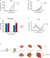
Analysis of Dand5 KO mESCs during cardiomyocyte differentiation. (A) Schematic representation of the cardiomyocytes’ differentiation using the hanging drop technique; (B) Relative Dand5 expression throughout cardiac differentiation. (C) Measurement of beating foci per KO and WT EBs. Results are expressed as the total number of beating foci with respect to the total number of plated EBs; (D) Whole-mount EB immunofluorescence analysis show positive expression of sarcomeric α-actinin on Dand5 KO and WT EBs differentiated for 6 days. DAPI was used as a positive control for nuclear staining (scale bars: white 200 μm; yellow 20 μm). (E) Analysis of FLK-1 and PDGFR-α expression by flow cytometry. All the results represent the mean ± SD of three independent biological experiments. Unpaired Student’s t-test was applied to compare the differences between WT and KO groups in each day of differentiation. Statistically significant results were considered when *p < 0.05, ***p < 0.001, and ****p < 0.0001.
FIGURE 3
FIGURE 3
Dand5 KO leads to increased cardiomyocyte proliferation (A) Relative mRNA expression of MHC in Dand5 KO and WT differentiated cells; (B) Relative cTNT expression throughout cardiac differentiation of Dand5 KO and WT cells; (C) Analysis of cardiomyocyte proliferation by labeling newly synthesized DNA using EdU incubation for 1.5 h. Relative fluorescence intensity of cells was analyzed by flow cytometry; (D) Relative mRNA expression of Ccnd1 gene in Dand5 KO and WT differentiated cells. (E) Summary of the results: the increased formation of cardiac progenitor cells and the increased proliferative capacity of Dand5 KO mESC-derived cardiomyocytes contribute to the observed higher number of derived cardiac cells. All the results represent the mean ± SD of three independent biological experiments. Unpaired Student’s t-test was applied to compare the differences between WT and KO groups in each day of differentiation. Statistically significant results were considered when *p < 0.05, **p < 0.01, and ****p < 0.0001.
FIGURE 4
FIGURE 4
Relative mRNA expression of Brachyury(T), Mesp-1, Fzd4, Bmp2, Nkx2.5, and Isl1 genes in Dand5 KO and WT differentiated cells. All the results represent the mean ± SD of three independent biological experiments. Unpaired Student’s t-test was applied to compare the differences between WT and KO groups in each day of differentiation. Statistically significant results were considered when *p < 0.05 and ****p < 0.0001.
FIGURE 5
FIGURE 5
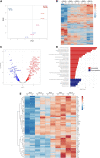
RNA-seq Analysis of EB differentiation up to day 10. (A) Principal Component Analysis (PCA) of WT and Dand5 KO samples from EB differentiation from mESC (D0) to day 10 of differentiation (D10); (B) Heat Map of all the genes commonly counted in all the samples. Profiles of expression of each gene were hierarchically clustered; (C) Volcano plot of genes differentially expressed at day 6 of EB differentiation. Genes with FDR < 0.01 were colored in red. Results are relative to WT data. (D) Pathway enrichment analysis of genes differentially expressed at day 6 of EB differentiation. Results are relative to WT data; (E) Heat Map of genes related with Heart Cardiomyopathy and Dilated Cardiomyopathy terms of KEGG. Profiles of expression of each gene were hierarchically clustered.

References

    1. Ames E. G., Lawson M. J., Mackey A. J., Holmes J. W. (2013). Sequencing of mRNA identifies re-expression of fetal splice variants in cardiac hypertrophy. J. Mol. Cell. Cardiol. 62 99–107. 10.1016/j.yjmcc.2013.05.004 - DOI - PMC - PubMed
    1. Araújo A. C., Marques S., Belo J. A. (2014). Targeted inactivation of cerberus like-2 leads to left ventricular cardiac hyperplasia and systolic dysfunction in the mouse. PLoS One 9:e102716. 10.1371/journal.pone.0102716 - DOI - PMC - PubMed
    1. Belo J. A., Bachiller D., Agius E., Kemp C., Borges A. C., Marques S., et al. (2000). Cerberus-like is a secreted BMP and nodal antagonist not essential for mouse development. Genesis 26 265–270. - PubMed
    1. Belo J. A., Marques S., Inácio J. M. (2017). The role of Cerl2 in the establishment of left-right asymmetries during axis formation and heart development. J. Cardiovasc. Dev. Dis. 4:23. 10.3390/jcdd4040023 - DOI - PMC - PubMed
    1. Belo J. A., Silva A. C., Borges A.-C., Filipe M., Bento M., Gonçalves L., et al. (2009). Generating asymmetries in the early vertebrate embryo: the role of the Cerberus-like family. Int. J. Dev. Biol. 53 1399–1407. 10.1387/ijdb.072297jb - DOI - PubMed