. 2021 Apr 13:9:647058.
doi: 10.3389/fcell.2021.647058. eCollection 2021.

Ecto-5'-Nucleotidase (CD73) Regulates the Survival of CD8+ T Cells

Affiliations

Ecto-5'-Nucleotidase (CD73) Regulates the Survival of CD8+ T Cells

Mariana V Rosemblatt et al. Front Cell Dev Biol. .

Abstract

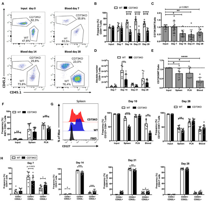
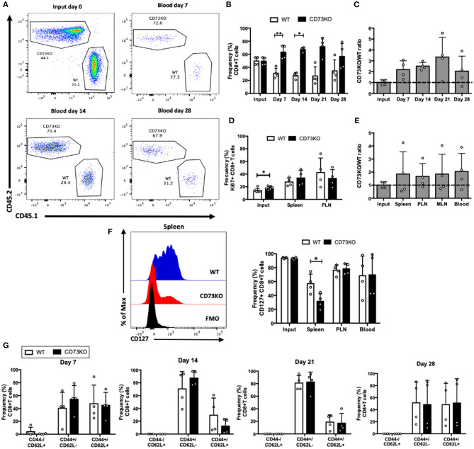
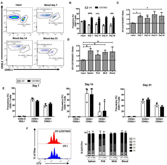
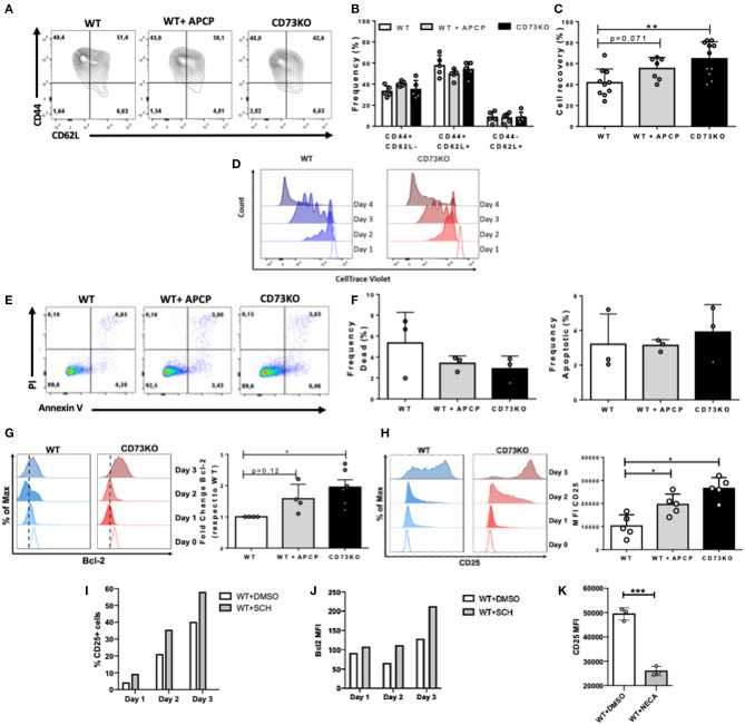
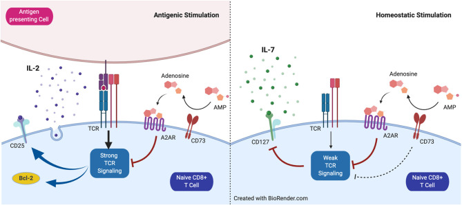
Ecto-5'-nucleotidase (CD73) is an enzyme present on the surface of tumor cells whose primary described function is the production of extracellular adenosine. Due to the immunosuppressive properties of adenosine, CD73 is being investigated as a target for new antitumor therapies. We and others have described that CD73 is present at the surface of different CD8+ T cell subsets. Nonetheless, there is limited information as to whether CD73 affects CD8+ T cell proliferation and survival. In this study, we assessed the impact of CD73 deficiency on CD8+ T cells by analyzing their proliferation and survival in antigenic and homeostatic conditions. Results obtained from adoptive transfer experiments demonstrate a paradoxical role of CD73. On one side, it favors the expression of interleukin-7 receptor α chain on CD8+ T cells and their homeostatic survival; on the other side, it reduces the survival of activated CD8+ T cells under antigenic stimulation. Also, upon in vitro antigenic stimulation, CD73 decreases the expression of interleukin-2 receptor α chain and the anti-apoptotic molecule Bcl-2, findings that may explain the reduced CD8+ T cell survival observed in this condition. These results indicate that CD73 has a dual effect on CD8+ T cells depending on whether they are subject to an antigenic or homeostatic stimulus, and thus, special attention should be given to these aspects when considering CD73 blockade in the design of novel antitumor therapies.

Keywords: CD127 (IL7 receptor); CD25; CD73/NT5E; CD8+ T cell; antigenic activation; homeostatic.


Conflict of interest statement

The authors declare that the research was conducted in the absence of any commercial or financial relationships that could be construed as a potential conflict of interest.

Figures

Figure 1
Figure 1
CD73 promotes in vivo homeostatic proliferation and CD127 expression in naïve CD8+ T cells. Naïve CD8+ T cells (CD8+/CD25-/CD44-/CD62L+) obtained from WT (CD45.1+) and CD73KO (CD45.2+) mice were co-transferred in Rag1–/– mice. Rag1–/– mice were bled at different days to analyze the frequency and phenotype of transferred cells. Mice were euthanized on day 28 to analyze the transferred cells in the spleen and peripheral lymph nodes (PLN). (A) FACS plots showing the input and frequency of transferred cells at days 7, 14, and 28 in the blood. (B) Frequency of transferred cells in the blood at input and days 7, 14, 21, and 28. (C) CD73KO/WT ratio at input and days 7, 14, 21, and 28 in blood. (D) Absolute numbers of transferred CD8+ T cells in the blood at days 7, 14, 21, and 28. (E) CD73KO/WT ratio at input and day 28 in the spleen, PLN, and the blood. (F) Frequency of Ki67+ cells (gated on transferred cells) at input, spleen and PLN at day 28. (G) Left, FACS histogram overlay depicting CD127 expression (gated on transferred cells) in the spleen at day 28. Right, frequency of CD127+ among transferred cells in the spleen, PLN and the blood at day 10 and 28. (H) Frequency of transferred cells expressing naïve (CD44-/CD62L+), effector memory (CD44+/CD62L–), and central memory (CD44+/CD62L+) phenotypes at days 7, 14, 21, and 28 in the blood. All data represent mean ± s.d. Data were analyzed by two-tailed unpaired Student's t-Test (B,D, F–H) or one-way ANOVA with Bonferroni post-hoc test (C,E). *p < 0.05; **p < 0.01; ***p < 0.005; ****p < 0.001.
Figure 2
Figure 2
CD73 has no effect on central memory CD8+ T cell differentiation and survival in homeostatic conditions. Central memory CD8+ T cells (CD8+/CD25–/CD44+/CD62L+) obtained from WT (CD45.1+) and CD73KO (CD45.2+) mice were co-transferred in Rag1–/– mice. Rag1–/– mice were bled at different time points to analyze the frequency and phenotype of transferred cells. Mice were euthanized on day 28 to analyze the transferred cells in the spleen, PLN, and mesenteric lymph node (MLN). (A) FACS plots showing the input and frequency of transferred cells at days 7, 14, and 28 in blood. (B) Frequency of transferred cells at input and in the blood at days 7, 14, 21, and 28. (C) CD73KO/WT ratio at input and days 7, 14, 21, and 28 in the blood. (D) Frequency of Ki67+ cells at input, spleen and PLN at day 28. (E) CD73KO/WT ratio at input and day 28 in the spleen, PLN, MLN, and the blood. (F) Left, FACS histogram overlay depicting CD127 expression in transferred cells in the spleen at day 28. Right, frequency of CD127+ transferred cells at input and in the spleen, PLN and the blood at day 28. (G) Frequency of transferred cells expressing naïve (CD44–/CD62L+), effector memory (CD44+/CD62L–), and central memory (CD44+/CD62L+) phenotypes at days 7, 14, 21, and 28 in the blood. All data represent mean ± s.d. Data were analyzed by two-tailed unpaired Student's t-Test (B), Mann–Whitney Test (D,F,G), or one-way ANOVA with Bonferroni post-hoc test (C,E). *p < 0.05; **p < 0.01.
Figure 3
Figure 3
CD73 reduces the frequency of CD8+ T cells following in vivo antigenic stimulation. Naïve CD8+ T cells (CD8+/CD25–/CD44-/CD62L+) obtained from OT-I (CD45.1+/CD45.2+) and OT-I/CD73KO (CD45.2+) mice were co-transferred in CD45.1+ mice. Twenty-four hours later, recipient mice were immunized i.p. with OVA protein (500 μg) plus Poly I:C (50 μg). Mice were bled at different time points to analyze the frequency and phenotype of transferred cells. Mice were euthanized on day 28 to analyze the transferred cells in the spleen, PLN, and MLN. (A) FACS plots showing the input and frequency of transferred cells at days 7, 14, and 21 in blood. (B) Frequency of transferred cells in the blood at input and days 7, 14, 21, and 28. (C) CD73KO/WT ratio at input and days 7, 14, 21, and 28 in the blood. (D) CD73KO/WT ratio at input and day 28 in the spleen, PLN, MLN, and the blood. (E) Frequency of transferred cells expressing naïve (CD44-/CD62L+), effector memory (CD44+/CD62L-), and central memory (CD44+/CD62L+) phenotypes at days 7, 14, and 21 in blood. (F) Histogram overlay for CTV dilution and bar graph depicting the frequency of transferred cells in each proliferation round in the spleen, PLN, MLN, and blood. Naïve CD8+ T cells (CD8+/CD24–/CD44–/CD62L+) obtained from OT-I (CD45.1+/CD45.2+) and OT-I/CD73KO (CD45.2+) mice were labeled with cell trace violet (CTV) and co-transferred in CD45.1+ mice. Twenty-four hours later, recipient mice were immunized i.p. with OVA protein (500 μg) plus LPS (25 μg). Mice were euthanized on day 4 to analyze the proliferation of transferred cells as assessed by CTV dilution in the spleen, PLN, MLN, and the blood. All data represent mean ± s.d. Data were analyzed by two-tailed unpaired Student's t-Test (B), Mann–Whitney Test (E,F), or one-way ANOVA with Bonferroni post-hoc test (C,D). *p < 0.05; **p < 0.01; ****p < 0.001.
Figure 4
Figure 4
CD73 reduces CD25 expression in CD8+ T cells following in vitro antigenic stimulation. Sorted naïve CD8+ T cells from WT and CD73KO mice were labeled with CTV and activated in vitro with antibodies against CD3/CD28 in the presence of IL-2 (10 ng/ml). Where indicated, APCP (50 μM), SCH58261 (5 μM), or NECA (1 μM) were added to the cultures. At different time points, cells were harvested and analyzed by FACS. (A) FACS plot depicting CD62L and CD44 expression in WT cells, APCP-treated WT cells, and CD73KO cells at day 4 following activation. (B) Frequency of cells expressing CD62L and CD44 for WT cells, APCP-treated WT cells, and CD73KO cells at day 4 following activation. (C) Percentage of cell recovery after 4 days of activation for WT cells, APCP-treated WT cells, and CD73KO cells. Percentage of cell recovery: (absolute number of cells obtained after culture/ absolute number of cells cultured at the start of the assay) × 100. (D) Histogram overlay depicting proliferation (as cell trace violet dilution) for WT and CD73KO cells at different time points following activation (representative of three independent experiments). (E) FACS plot showing PI/Annexin V staining following 4 days of activation for WT cells, APCP-treated WT cells, and CD73KO cells. (F) Frequency of dead and apoptotic cells recovered after 4 days of activation for WT cells, APCP-treated WT cells, and CD73KO cells. (G) Left, histogram overlay showing Bcl-2 expression at different time points for WT and CD73KO cells. Right, Bcl-2 expression fold-change (relative to WT cells) in CD73KO cells and APCP-treated WT cells after 3 days of activation. (H) Left, histogram overlay depicting CD25 expression at different time points for WT and CD73KO cells. Right, bar graph showing CD25 mean fluorescence intensity (MFI) following 3 days of activation for WT, CD73KO and APCP-treated cells. (I) Percentage of CD25+ cells for WT cells at different time points following activation in the presence of SCH58261 or its vehicle control (DMSO). (J) Bcl2 MFI for WT cells at different time points following activation in the presence of SCH58261 or its vehicle control (DMSO). (K) CD25 MFI for WT cells at day 4 following activation in the presence of NECA or its vehicle control (DMSO). All data represent mean s.d. Data were analyzed by the Kruskal-Wallis test with Dunn's post-hoc test (B,C,F–H) and by two-tailed unpaired Student's t-Test (K) *p < 0.05; **p < 0.01, ***p < 0.005.
Figure 5
Figure 5
Proposed model for the role of CD73 in homeostatic and antigenic stimulation of naïve CD8+ T cells. CD73 reduces signaling mediated by the T cell receptor (TCR) and restrains CD25 and Bcl-2 expression under antigenic stimulation. Under homeostatic conditions, CD73 prevents CD127 downregulation mediated by weak TCR stimulation.

Similar articles

Cited by

References

    1. Akashi K., Kondo M., von Freeden-Jeffry U., Murray R., Weissman I. L. (1997). Bcl-2 rescues T lymphopoiesis in interleukin-7 receptor-deficient mice. Cell 89, 1033–1041. 10.1016/S0092-8674(00)80291-3 - DOI - PubMed
    1. Allard D., Allard B., Gaudreau P. O., Chrobak P., Stagg J. (2016). CD73-adenosine: a next-generation target in immuno-oncology. Immunotherapy 8, 145–163. 10.2217/imt.15.106 - DOI - PubMed
    1. Antonioli L., Novitskiy S. V., Sachsenmeier K. F., Fornai M., Blandizzi C., Haskó G. (2017). Switching off CD73: a way to boost the activity of conventional and targeted antineoplastic therapies. Drug Discov. Today 22, 1686–1696. 10.1016/j.drudis.2017.06.005 - DOI - PubMed
    1. Barata J. T., Durum S. K., Seddon B. (2019). Flip the coin: IL-7 and IL-7R in health and disease. Nat. Immunol. 20, 1584–1593. 10.1038/s41590-019-0479-x - DOI - PubMed
    1. Becker T. C., Wherry E. J., Boone D., Murali-Krishna K., Antia R., Ma A., et al. . (2002). Interleukin 15 is required for proliferative renewal of virus-specific memory CD8 T cells. J. Exp. Med. 195, 1541–1548. 10.1084/jem.20020369 - DOI - PMC - PubMed