Review
. 2021 Apr 7:2021:5528006.
doi: 10.1155/2021/5528006. eCollection 2021.

The Use of Radial Artery for CABG: An Update

Affiliations
Review

The Use of Radial Artery for CABG: An Update

Francesco Nappi et al. Biomed Res Int. .

Abstract

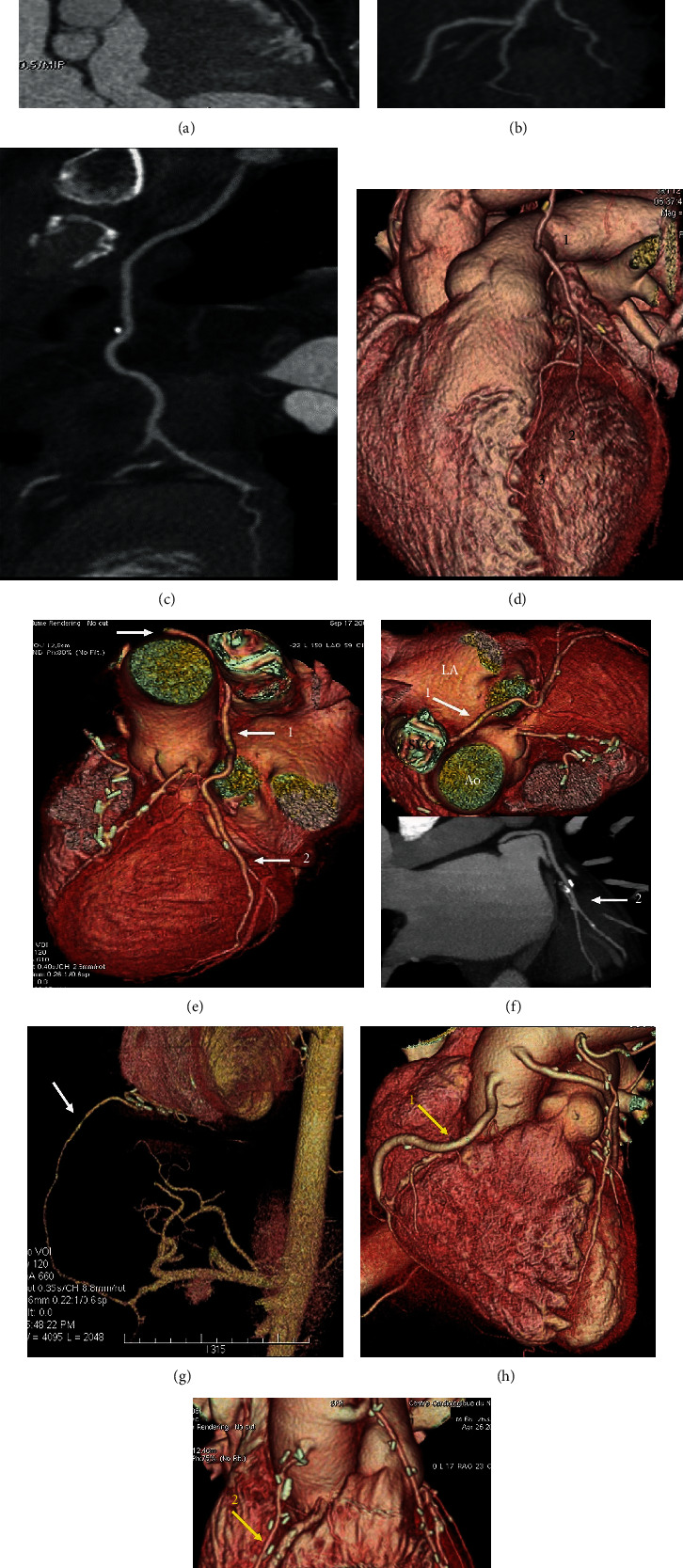
We used the radial artery as a second target conduit for coronary artery bypass grafting since 1971. However, randomized clinical studies have demonstrated differences in clinical outcomes between the radial artery and other grafts because these trials are underpowered. As we proceed toward 50 years of experience with radial artery grafting, we examined the literature to define the best second-best target vessel for coronary artery bypass grafting. The literature was reviewed with emphasis, and a large number of randomized controlled trials, propensity-matched observational series, and meta-analyses were identified with a large patient population who received arterial conduit and saphenous vein grafts. The radial artery has been shown to be effective and safe when used as a second target conduit for coronary artery bypass grafting. Results and patency rates were superior to those for saphenous vein grafting. It has also been shown that the radial artery is a safe and effective graft as a third conduit into the territory of the artery right coronary artery. However, there is little evidence based on a few comparable series limiting the use of the gastroepiploic artery. In its fifth decade of use, we can finally deduced that the aorto-to-coronary radial bypass graft is the conduit of choice for coronary operations after the left internal thoracic artery to the left anterior descending artery.


Conflict of interest statement

All authors declare no conflicts of interest.

Figures

Figure 1
Figure 1
(a–i) Postprocessing of CT angiography of CABG by mean of volume rendering and 2D curved imaging with automatic tracking. (a–c) show 2D curved imaging of CABG with automatic tracking. (a) LITA anastomosed on LAD. (b) LITA sequential grafting on LAD and first diagonal branch. (c) RITA anastomosed on first obtuse branch. (d-i) Volume rendering imaging of CABG. (d) 1-LITA grafted on 2-Diag branch and 3-LAD. (e, f) RITA grafted on CCA. White arrow (1) RITA runs between the aorta and LA, (2) distal grafting on first obtuse branch. (g–i) Comparison between second target conduit on RCA. (g) White arrow gastroepiploic anastomosed to the PDA. (h, i) Yellow arrow (1) SVG and (2) RAG. Note that the venous graft size is greater than the arterial graft size. CABG: coronary artery bypass grafting; LITA: left internal thoracic artery; LAD: left anterior descending; RITA: right internal artery; CCA: circonflexe coronary artery; LA: left appendix; RCA: right coronary artery; PDA: posterior descending artery; SVG: saphen vein graft; RAG: radial artery graft.
Figure 2
Figure 2
(a) Asymptomatic brachial artery occlusion fortuitously discovered on a postoperative coronary angiogram in a patient having undergone radial artery grafting. This case belongs to the early series. Nowadays, preoperative echo-Doppler of the upper-limb would have revealed the lesion, and RA would not have been used. (b, c) Media calcinosis of the radial artery. (b) Nonobstructive mediacalcinosis in a diabetic patient; (c) atheromatous occlusion with intraluminal thrombus. (d) Traumatic injury for transradial artery coronary intervention. Note that the size of the sheath (arrow) equals that of the vessel lumen.
Figure 3
Figure 3
(a–f) CT angiographic control of RA used as second target conduit on second obtuse branch of LCC and PDA. (a) Volume rendering imaging of RA (yellow arrow) running at the level of Theil sinus transversus. (b–f) 2D curved imaging. (b) Course of the radial artery; (c) stenosis of the body of the RA graft. (d) Red arrow shows the metallic clip while yellow arrow highlights a fibrous plaque that are contiguous. (e) Yellow arrow shows a calcific plaque of RA conduit. (f) Note in the blue circle the stenosis of RA due to a fibrous plaque; red arrow shows a remodeling plaque. (LCC2). RA: radial artery; LCC: left coronary circumflex; LCC1: first obtuse branch; LCC2: second obtuse branch; RCA: right coronary artery.

References

    1. Carpentier A., Guermonprez J. L., Deloche A., Frechette C., DuBost C. The aorta-to-coronary radial artery bypass graft: a technique avoiding pathological changes in grafts. The Annals of Thoracic Surgery. 1973;16(2):111–121. doi: 10.1016/S0003-4975(10)65825-0. - DOI - PubMed
    1. Geha A. S., Krone R. J., McCormick J. R., Baue A. E. Selection of coronary bypass: Anatomic, physiological, and angiographic considerations of vein and mammary artery grafts. The Journal of Thoracic and Cardiovascular Surgery. 1975;70(3):414–431. doi: 10.1016/S0022-5223(19)40315-2. - DOI - PubMed
    1. Acar C., Jebara V. A., Portoghese M., et al. Revival of the radial artery for coronary artery bypass grafting. The Annals of Thoracic Surgery. 1992;54(4):652–660. doi: 10.1016/0003-4975(92)91007-V. - DOI - PubMed
    1. Acar C., Ramsheyi A., Pagny J.-Y., et al. The radial artery for coronary artery bypass grafting: clinical and angiographic results at five years. The Journal of Thoracic and Cardiovascular Surgery. 1998;116(6):981–989. doi: 10.1016/S0022-5223(98)70050-9. - DOI - PubMed
    1. Achouh P., Boutekadjirt R., Toledano D., et al. Long-term (5- to 20-year) patency of the radial artery for coronary bypass grafting. The Journal of Thoracic and Cardiovascular Surgery. 2010;140(1):73–79.e2. doi: 10.1016/j.jtcvs.2009.09.032. - DOI - PubMed