Review
. 2021 Jun 4;48(3-4):kuab005.
doi: 10.1093/jimb/kuab005.

Mechanisms of action of ribosomally synthesized and posttranslationally modified peptides (RiPPs)

Affiliations
Review

Mechanisms of action of ribosomally synthesized and posttranslationally modified peptides (RiPPs)

Li Cao et al. J Ind Microbiol Biotechnol. .

Abstract

Natural products remain a critical source of medicines and drug leads. One of the most rapidly growing superclasses of natural products is RiPPs: ribosomally synthesized and posttranslationally modified peptides. RiPPs have rich and diverse bioactivities. This review highlights examples of the molecular mechanisms of action that underly those bioactivities. Particular emphasis is placed on RiPP/target interactions for which there is structural information. This detailed mechanism of action work is critical toward the development of RiPPs as therapeutics and can also be used to prioritize hits in RiPP genome mining studies.

Keywords: Mechanism of action; Natural products; RiPPs.


Figures

Fig. 1.
Fig. 1.
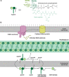
The varied targets of bioactive RiPPs. Members of the RiPPs superfamily exert antibiotic and/or cytotoxic activity via inhibition of nearly all major cellular processes. Many RiPPs are membrane-active, compromising the permeability barrier of the cytoplasmic membrane in both Gram-negative and Gram-positive bacteria (only a Gram-negative cell is shown for simplicity's sake). Other RiPPs active at the cell envelope disrupt outer membrane biogenesis in Gram-negative bacteria and peptidoglycan (cell wall) biosynthesis. RiPPs also engage cytoplasmic targets disrupting the central processes of DNA replication, transcription, and translation. OM: outer membrane, IM: inner membrane.
Fig. 2.
Fig. 2.
The bacterial cell envelope is a common target for RiPPs. (A) Cartoon and chemical structure of lipid II, the precursor to peptidoglycan. The stem peptides of lipid II from different bacteria vary at the positions indicated by R1, R2, and R3. GlcNAc: N-acetylglucosamine, MurNAc: N-acetylmuramic acid. (B) Schematic of cell envelope biosynthesis processes disrupted by RiPPs. Both lanthipeptides and the lasso peptide siamycin-I bind to lipid II, disrupting peptidoglycan biosynthesis. The recently discovered RiPP darobactin inhibits BamA, disrupting the assembly of outer membrane proteins in Gram-negative bacteria. BAM: β-barrel assembly machinery.
Fig. 3.
Fig. 3.
Structure and mechanism of action of the lanthipeptide nisin. (A) Representation of nisin structure. Dhb: dehydrobutyrine, Dha: dehydroalanine, Abu: aminobutyric acid. The thioether linkage between Ala and Ala is referred to as a lanthionine moiety while the linkage between Abu and Ala is a methyllanthionine linkage. (B) NMR structure (PDB file 1WCO) of nisin bound to a lipid II analog. Nisin is shown as a space-filling model and the lipid II analog is shown as sticks. The N-terminal (methyl)lanthionine rings (space-filling tan) of nisin envelop the pyrophosphate moiety (magenta and cyan sticks) of lipid II.
Fig. 4.
Fig. 4.
The β-spiral structure of polytheonamide B in organic solvent to mimic the membrane environment (PDB file 2RQO). Left: View showing the length of polytheonamide B to be roughly the same thickness as a plasma membrane. Note the density of polar residues at the C-terminus of the structure. Right: The interior of polytheonamide B is polar, allowing for the passage of cations such as potassium. Polytheonamide B is a potent cytotoxin, functioning as an ion channel.
Fig. 5.
Fig. 5.
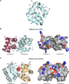
Structure of a synthetic analog of pediocin PA-1, a membrane-active bacteriocin. This compact structure is formed via two disulfide bonds, which are highlighted in orange. The N-terminal region of pediocin PA-1 is cationic, allowing for electrostatic interactions with bacterial membrane lipid head groups, while its C-terminal region is hydrophobic, allowing for pore formation in membranes. Figure drawn from PDB file 5UKZ.
Fig. 6.
Fig. 6.
Structure of the membrane-active bacteriocin enterocin AS-48. (A) Enterocin AS-48 is cyclized between its N- and C-termini and highly helical. Sidechains of negatively and positively charged amino acids are clustered together in helices 4 and 5 and are shown as sticks (PDB file 1O82). (B and C) Two distinct dimeric forms of enterocin AS-48 are found within the crystal structure. Dimeric form I (DF-I) shows the hydrophobic side chains of helices 1 and 2 in gray. Dimeric form II (DF-II) includes a sulfate ion at the protomer–protomer interface.
Fig. 7.
Fig. 7.
The structure of sactipeptide subtilosin A. (A) The 35 aa subtilosin A is cyclized via a head-to-tail peptide bond and three unusual thioether linkages between the side chain of cysteine and the α-carbon of a distal amino acid. (B) The solution structure of subtilosin A in methanol (PDB file 1PXQ) shows a compact hairpin-like structure. Acidic (D16, D21, E23) and basic (K2) residues are concentrated at different ends of the folded peptide. Residues participating in thioether linkages are shown as gold sticks.
Fig. 8.
Fig. 8.
Chemical structure of the heptapeptide RiPP darobactin. Linkages between sidechains are colored in blue.
Fig. 9.
Fig. 9.
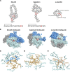
RiPP inhibitors of RNA polymerase (RNAP). (A) Schematics of microcin J25 (MccJ25), capistruin, and α-amanitin. ILX: 4,5-dihydroxyisoleucine, TRX: 6-hydroxytryptophan, CSX: S-oxy cysteine, HYP: HYP: 4-hydroxyproline. Note the tryptathionine linkage between 6-hydroxytryptophan and S-oxy cysteine. (B) From left to right, crystal structures of E. coli RNAP showing the binding site of MccJ25, E. coli RNAP showing the binding site of capistruin, and yeast RNA polymerase II (Pol II) showing the binding site for α-amanitin. Residues in RNAP or Pol II that directly contact these RiPPs are colored red. (C) Zoomed-in view of MccJ25 (gold/blue) bound to RNAP (cyan). Key amino acids for this interaction are labeled. Dashed lines are H-bonds (yellow). (D) As in part C, but for the RNAP–capistruin interaction. (E) Zoomed-in view of the α-amanitin-Pol II binding site with key interacting residues highlighted. HYP: hydroxyproline, ILX: dihydroxyisoleucine, TRX: hydroxytryptophan. Drawn from PDB files 6N60 (MccJ25), 6N61 (capistruin), and 3CQZ (α-amanitin).
Fig. 10.
Fig. 10.
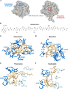
Examples of RiPPs that inhibit translation. (A) Crystal structures of bacterial ribosomes with thiopeptide thiostrepton binding site (PDB file 3CF5) colored in blue and linear azo(lin)e peptide (LAP) phazolicin binding site colored in red (PDB file 6U48). (B) Chemical structure of the LAP klebsazolicin. (C) Key interactions between klebsazolicin (gold) and 23S rRNA (blue), drawn from PDB file 5W4K. Dashed lines are H-bonds (yellow), and double-sided arrows represent π stacking. (D): As in part C, but for the phazolicin:ribosome interaction, drawn from PDB file 6U48. (E) Key interactions between thiostrepton (gold), 23S rRNA (blue), and ribosomal protein L11 (cyan) drawn from PDB file 3CF5. (F) As in part E, but for the nosiheptide:ribosome interaction, drawn from PDB file 2ZJP. THZ: thiazole, OXZ: oxazole, QA: quinaldic acid.
Fig. 11.
Fig. 11.
Interaction of thiopeptide GE2270A with elongation factor EF-Tu. (A) Crystal structure of the EF-Tu: GE2270A complex, drawn from PDB file 1D8T. (B) Close up view of the EF-Tu (cyan):GE2270A (magenta) complex. Amino acids from EF-Tu making direct contact with the thiopeptide are labeled. Atoms from GE2270A making direct contact are labeled. Dashed lines are H-bonds (yellow) or a salt bridge (blue) between R223 and E259 that accounts for the high stability of the GE2270A:EF-Tu complex.
Fig. 12.
Fig. 12.
Cellular uptake of lasso peptide microcin J25 (MccJ25) is mediated by the TonB-dependent transporter FhuA. (A) The canonical role of FhuA, an outer membrane (OM) receptor, is transport of iron-siderophore complexes across the OM into the periplasm (P). This energy-dependent process is mediated by the TonB/ExbB/ExbD inner membrane (IM) protein complex and driven by the proton motive force (PMF). (B) Crystal structure of MccJ25 bound to the extracellular face of FhuA. (C) Zoomed-in view of the FhuA:MccJ25 complex showing close contacts between the cork domain (gold) and β-barrel (cyan) of FhuA with MccJ25 (magenta). Dashed lines are H-bonds (yellow). All structure figures drawn from PDB file 4CU4.

Similar articles

Cited by

References

    1. Allgaier H., Jung G., Werner R. G., Schneider U., Zähner H. (1985). Elucidation of the structure of epidermin, a ribosomally synthesized, tetracyclic heterodetic polypeptide antibiotic. Angewandte Chemie (International Ed. in English), 24, 1051–1053. 10.1002/anie.198510511. - DOI
    1. Alvarez-Sieiro P., Montalbán-López M., Mu D., Kuipers O. P. (2016). Bacteriocins of lactic acid bacteria: Extending the family. Applied Microbiology and Biotechnology, 100, 2939–2951. 10.1007/s00253-016-7343-9. - DOI - PMC - PubMed
    1. Andersen O. S., Koeppe R. E. (2007). Bilayer thickness and membrane protein function: An energetic perspective. Annual Review of Biophysics and Biomolecular Structure, 36, 107–130. 10.1146/annurev.biophys.36.040306.132643. - DOI - PubMed
    1. Anderssen E. L., Diep D. B., Nes I. F., Eijsink V. G. H., Nissen-Meyer J. (1998). Antagonistic activity of Lactobacillus plantarum C11: Two new two- peptide bacteriocins, plantaricins EF and JK, and the induction factor plantaricin A. Applied and Environmental Microbiology, 64, 2269–2272. 10.1128/aem.64.6.2269-2272.1998. - DOI - PMC - PubMed
    1. Arioli V., Berti M., Silvestri L. G. (1976). Gardimycin, a new antibiotic from Actionoplanes. III. Biological properties. Journal of Antibiotics (Tokyo), 29, 511–515. 10.7164/antibiotics.29.511. - DOI - PubMed